A 54-year-old female presents to a chiropractic clinic with a chief complaint of a severe and debilitating exacerbation of chronic neck pain and stiffness. The patient’s narrative is one that is likely familiar to many practitioners. For years, she has navigated a baseline of manageable, intermittent neck stiffness, which she had pragmatically attributed to the combined effects of aging and the postural demands of her career as an accountant. This chronic condition was characterized by a low-grade ache at the base of the neck and a noticeable, but not limiting, loss of flexibility.
However, the clinical picture changed dramatically three weeks prior to her presentation. While stopped at an intersection, her vehicle was struck from behind in a minor, low-speed motor vehicle collision. Despite minimal damage to her car, the incident provoked an immediate and intense inflammatory response in her cervical spine. Her chronic, 2/10 stiffness escalated to a constant, sharp 7/10 pain. The once-manageable loss of motion has now become a functional impairment; she reports significant difficulty with daily activities such as checking her blind spot while driving, looking up at a high shelf, and even finding a comfortable position for sleep. The failure of her symptoms to improve with rest and standard over-the-counter anti-inflammatory medications prompted her to seek a more thorough evaluation and a definitive diagnosis.
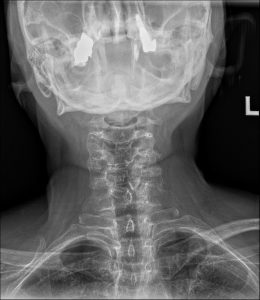
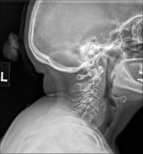
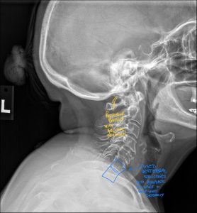
To investigate the underlying cause of her disproportionate response to a minor trauma, a standard three-view cervical spine radiographic series was ordered. The images provided a clear and immediate explanation for her clinical presentation.
On the lateral cervical view, the most striking finding is a congenital failure of segmentation involving the C6 and C7 vertebral bodies, their posterior articular pillars, and the corresponding spinous processes. This osseous fusion creates a single, solid block. At the anterior margin of this C6-C7 block, a distinct concavity or indentation is present at the level of the rudimentary disc. This feature, known as the “wasp-waist” sign, is a pathognomonic indicator of the congenital nature of the fusion.
The diagnostic story, however, does not end with the block vertebra itself. The true source of her pain and dysfunction is revealed at the adjacent motion segment above the block. The C5-C6 level demonstrates moderate to advanced degenerative changes. There is significant anterior osteophyte formation, marked narrowing of the intervertebral disc space, and notable facet arthrosis with sclerosis of the articular surfaces. These degenerative findings are far more advanced than would be typically expected for a 54-year-old, pointing to a long history of abnormal biomechanical stress. The radiology report clearly delineated these two related but distinct findings.
1. Primary Diagnosis: Congenital Block Vertebra at C6-C7. This is the underlying structural anomaly that has been present since birth.
2. Secondary Diagnosis: Advanced Secondary Degenerative Spondylosis and Arthrosis at the Hypermobile C5-C6 Segment, presenting with an acute traumatic exacerbation. This is the acquired, pathological consequence of the primary anomaly and the direct source of the patient’s clinical symptoms.
To truly appreciate the nature of a block vertebra, it is helpful to revisit the remarkable process of spinal development in the embryo. During the third week of gestation, the paraxial mesoderm organizes into segmented blocks of tissue called somites. These somites differentiate into sclerotomes, which are the precursors to the vertebrae.
A crucial phase in this process is “resegmentation.” Each sclerotome splits into a cranial (top) half and a caudal (bottom) half. The caudal half of one sclerotome then fuses with the cranial half of the sclerotome immediately below it to form a single vertebral body. This brilliant process ensures that the intersegmental arteries pass over the middle of the vertebral bodies and, more importantly, that the myotomes (muscle precursors) bridge the intervertebral disc spaces, allowing for spinal movement.
A congenital block vertebra represents a localized failure of this resegmentation process. For reasons not fully understood, the cleft that should form between two adjacent sclerotome halves fails to develop. As a result, the C6 and C7 vertebral precursors never separate, remaining a single cartilaginous block that eventually ossifies into the fused vertebra seen in this patient. This embryological misstep is the root cause of the entire clinical picture.
The human cervical spine is an engineering marvel, designed to balance the seemingly contradictory demands of stability (to protect the spinal cord) and mobility (to allow for a vast range of head movements). The normal spine dissipates forces through a coordinated system of seven mobile vertebrae.
When a segment like C6-C7 is fused, it effectively removes a shock absorber from the system. The fused block becomes a rigid, unyielding lever arm. Consequently, the kinetic forces that would normally be distributed across the C6-C7 disc and facets are shunted to the adjacent mobile segments. The C5-C6 and C7-T1 levels are forced to compensate for the lost motion, entering a state of chronic hypermobility.
This hypermobility is not benign. The C5-C6 disc and facets are now subjected to decades of excessive shear and compressive forces, moving beyond their intended physiological range with every nod, turn, and tilt of the head. This overload accelerates the natural degenerative cascade. The annulus fibrosus of the disc weakens, the nucleus pulposus dehydrates, and the articular cartilage of the facet joints wears thin. The body’s attempt to stabilize the overworked segment results in the formation of osteophytes (bone spurs) and ligamentous thickening—the very degenerative changes seen on this patient’s radiographs. The block vertebra creates a “stress riser” at the C5-C6 level, making it the weak link in the kinetic chain and exquisitely vulnerable to injury from trauma.
A key takeaway from this case is the importance of a thorough diagnostic workup. A patient presenting with neck pain after a minor MVC could easily be diagnosed with a simple “cervical sprain/strain.” However, several clues in this patient’s history pointed toward a more complex underlying issue:
Disproportionate Symptoms: The severity of her pain was inconsistent with the minor nature of the trauma.
Chronic History: Her pre-existing stiffness suggested an underlying structural or mechanical problem.
Lack of Resolution: Failure to respond to standard conservative home care is a red flag.
These clinical indicators prompted the decision to obtain diagnostic imaging. While MRI and CT have their roles, plain film radiography remains the most cost-effective and efficient method for identifying osseous anomalies like a block vertebra. It provided a definitive answer, shifting the entire course of case management. This highlights a core tenet of modern chiropractic practice: diagnose first, then treat.
Receiving a radiology report from a qualified radiologist is a critical step, but understanding its nuances is what empowers the clinician. A report from a board-certified chiropractic radiologist, or DACBR, is particularly valuable in these cases. A DACBR is trained not only to identify pathology but also to understand its direct biomechanical and clinical implications for manual therapy.
A high-quality radiology report in a case like this, provided by expert Diagnostic Imaging Consultants, will contain several key elements:
1. Identification of the Anomaly: Clearly states the presence of the C6-C7 congenital block vertebra.
2. Confirmation of Congenital Nature: Mentions the pathognomonic signs, like the “wasp-waist” deformity and rudimentary disc, differentiating it from an acquired (e.g., surgical) fusion.
3. Detailed Evaluation of Adjacent Segments: This is the most clinically relevant section. The report will meticulously describe the degenerative changes at C5-C6, grading their severity. This confirms the presence of adjacent segment disease and identifies the likely pain generator.
4. Clinical Correlation and Recommendations: The best reports bridge the gap between imaging and practice. A DACBR may include a comment such as, “The findings are consistent with adjacent segment hypermobility syndrome, which is the likely cause of the patient’s symptomatology. Mechanical stress at the C6-C7 fused segment should be avoided.”
This level of detail transforms the radiology report from a simple list of findings into a practical roadmap for creating a safe and effective treatment plan. It underscores the value of using specialized Diagnostic Imaging Consultants who understand the specific needs of the chiropractic physician.
The management of an uncomplicated fracture of the proximal phalanx of the fourth toe is primarily conservative.
Referral to Specialists: Referral to a podiatrist or orthopedic surgeon is required if the X-ray, interpreted in the chiropractic radiology report, shows a fracture that is significantly displaced, unstable, or involves the joint surface with a large step-off. These factors increase the risk of a poor outcome and may require manual reduction or surgical fixation.
The Chiropractic Radiologist’s Role: The DACBR ensures the treating chiropractor has the most accurate diagnosis from the imaging. They provide crucial consultation to confirm that the fracture is indeed stable enough for conservative management, thus preventing a malunion that could cause chronic foot problems.
Every day, chiropractors face the same frustration: imaging reports that miss what matters. General radiologists weren’t trained in your world; they don’t understand subluxations, joint dysfunction, or the biomechanical findings that drive your treatment decisions.
The result? Delayed care. Uncertain patients. Cases that stall when they should be progressing.
The Kinetic Radiology Difference: Chiropractors Reading for Chiropractors
Our board-certified DACBRs aren’t just radiologists. We’re chiropractors who chose to specialize in musculoskeletal imaging. We speak your language because we’ve stood where you stand.
Reports You Can Act On Immediately – No vague findings. No irrelevant details. Just the specific insights that guide your next adjustment, your treatment plan, and your patient conversations.
Same-Day Turnaround – Your patients don’t want to wait days wondering what’s wrong. Neither should you. Get clarity fast so care never stalls.
Documentation That Protects Your Practice – Whether it’s insurance requirements, legal protection, or patient records, our reports give you the clinical backing you need.
Confidence That Builds Your Reputation – When patients see you consulting with specialized radiologists, they recognize your commitment to excellence. That trust turns into loyalty, referrals, and five-star reviews.
Think about the last complex case you handled. Did the radiology report actually help you—or did you have to fill in the gaps yourself?
Now imagine having a DACBR partner who catches the subtle findings, flags the red flags, and gives you confidence in every diagnosis.
No commitment. No risk. Just submit your next challenging case and experience what specialized chiropractic radiology can do for your clinical confidence and patient outcomes.
Questions? Call us at 321 325 0096 or email at support@kineticradiology.com
No, the block itself is not dangerous. The main concern is that it can cause extra “wear and tear” on the joints next to it over time.
The fused block is actually very strong and stable. The issue is that because that one spot can’t move, the joints directly above and below it have to move extra to make up for it. Over many years, this extra work can lead to arthritis or soreness in those neighboring joints. The goal of chiropractic care and physical therapy is to manage that extra stress to keep you feeling great.
The block vertebra made your neck more vulnerable, and a recent event like an injury, poor posture, or even just aging likely triggered the pain.
For years, your body did a great job of compensating for the fused segment. However, an event like a minor car accident, a period of prolonged desk work, or the natural aging process can overwhelm that compensation. The pain you feel is typically coming from the overworked joints next to the block, which have finally become inflamed or irritated.
It creates a rigid lever arm, inducing compensatory hypermobility and accelerated spondylosis at the adjacent segments.
The fused segment acts as a point of stress shielding. All sagittal and rotational forces that would normally be attenuated at that level are shunted to the adjacent intervertebral junctions. This results in chronic hypermobility, predisposing the facet joints to arthrosis and the intervertebral disc to accelerated degeneration. This can also create hypomobility and muscular hypertonicity in segments further up or down the kinetic chain.
A congenital block has a “wasp-waist” sign and a rudimentary disc, while an acquired fusion (surgical or post-traumatic) has a uniform anterior margin and often shows evidence of hardware.
A high-quality radiology report from a DACBR will make this distinction. Key findings for a congenital block include: 1) the pathognomonic anterior concavity (wasp-waist), 2) a remnant of the disc space, and 3) frequent fusion of the posterior elements. An acquired fusion typically has a “blocky” appearance with a straight anterior border and lacks these congenital features.
The prognosis is generally poorer, with an increased likelihood of a prolonged recovery period and a higher risk of chronic symptoms.
The presence of a block vertebra creates a stress riser, meaning the hypermobile adjacent segments are likely to sustain more significant soft tissue damage in a whiplash event. This predisposes the patient to a more severe inflammatory response and a longer, more complicated recovery. Clinicians should set realistic expectations with the patient regarding their healing timeline.
Partnering with a DACBR teleradiology service provides more than just a second opinion; it offers a significant return on investment:
Speed: Get expert reports in hours, not days.
Expertise: Access board-certified specialists without having to hire them.
Convenience: The entire process is handled online from your office.
Clarity: Receive clear, concise reports that are clinically relevant to chiropractic care, not generic medical reports.
Posted onTrustindex verifies that the original source of the review is Google. Kinetic radiology has been an absolute game changer in speed of reports and detailed reports. Any other doctors I send my reports to are amazed at the detail and the pathology that gets picked up. This is my one and only radiologist group, im thrilled.Posted onTrustindex verifies that the original source of the review is Google. Rishi provides an outstanding service—fast, reliable, and incredibly reassuring. He’s quick to respond, efficient in his work, and always takes the time to address any concerns with clarity and professionalism. I highly recommend his services to anyone looking for a dependable DACBR.Posted onTrustindex verifies that the original source of the review is Google. Prompt efficient service that is thorough and clear. Spinal information is top notch and I've had patients discover kidney stones and possible issues with a hip joint replacement loosening as incidental findings that supported both me and the patient above expectations.Posted onTrustindex verifies that the original source of the review is Google. Quick, accurate, and easy to work with. My new radiology team!Posted onTrustindex verifies that the original source of the review is Google. Excellent, timely reads. Invaluable for CBCTPosted onTrustindex verifies that the original source of the review is Google. Best turnaround time and thorough reports out of any radiologist I’ve seen or worked with!Posted onTrustindex verifies that the original source of the review is Google. Very detailed reports and quick service. Highly recommendedPosted onTrustindex verifies that the original source of the review is Google. Fast turn around time for the radiology reports! Thank you for making this process as seamless as possible!Posted onTrustindex verifies that the original source of the review is Google. I am a NUCCA chiropractor located in Wauankee Wisconsin and I can tell you Dr. Rishi is the only radiologist I’d work with. Sure there are many others in my area but when you want the best you go to the best. He is very easy to work with and always fast to respond and report. 100% recommend.Posted onTrustindex verifies that the original source of the review is Google. Kinetic Radiology is great! They were able to read and get a report written immediately. They are my go to company for any and all images that I need read!Load more
We service all 50 U.S. States, including the following States and Cities listed below.
Copyright 2024 Kinetic Radiology All Rights Reserved
Website Privacy | Terms of UseReceive timely resources to keep you and your practice on the cutting edge of Chiropractic Radiology.
Copyright 2024 Kinetic Radiology
All Rights Reserved
Receive timely resources to keep you and your practice on the cutting edge of Chiropractic Radiology.