Is that “hot, swollen ankle” in your diabetic patient truly cellulitis, or is it the “quiet destroyer”? Charcot neuroarthropathy is a master mimic, a “can’t-miss” diagnosis that often leads to catastrophic collapse and amputation when misidentified. Read our clinical guide for healthcare professionals on the key imaging findings, differential diagnosis, and urgent management protocols to confidently identify and manage acute Charcot.
A 58-year-old male with a 15-year history of Type 2 diabetes mellitus presents with a chief complaint of a “swollen ankle” for three weeks. He denies any specific, significant trauma but vaguely recalls “twisting it a bit” while stepping off a curb. His primary concern is that the swelling is not resolving, and he can no longer fit his foot into a shoe.
He has already completed a 10-day course of cephalexin prescribed by his primary care provider for suspected cellulitis, with no improvement.
Clinically, the right ankle is profoundly edematous with significant, non-pitting edema and diffuse erythema. The skin is warm to the touch, measuring 4°C warmer than the contralateral asymptomatic side. The most striking finding, however, is the patient’s reported pain: he rates it a “2/10,” describing it only as a “dull ache.”
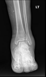
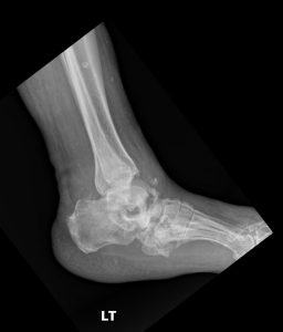
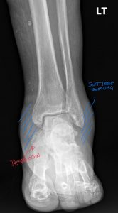
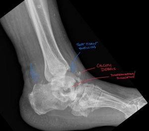
Acute Charcot Neuroarthropathy of the ankle.
In the daily gauntlet of clinical practice, we are detectives, trained to follow a trail of clues. Pain, swelling, redness, loss of function—these are the signposts that guide our diagnostic reasoning.
But what if the clues are lying? What if the body’s most reliable informant—pain—has been silenced?
Welcome to the diagnostic minefield of Charcot neuroarthropathy (CN). This is not a subtle condition. It is a violent, progressive, and destructive joint disease that moves with shocking speed. It is a “fire in the house” that consumes bone and cartilage, often in a matter of weeks. And it is happening right under our noses.
The hard truth is that Charcot neuroarthropathy is not a “zebra.” In the setting of our global diabetes epidemic, it is a tragically common horse that we are still misidentifying. We mistake it for cellulitis, for gout, or for a simple sprain.
And with every misdiagnosis, with every week the patient is allowed to walk on it, we are not just missing a call—we are co-signing an amputation. The 5-year mortality rate following a major lower-extremity amputation rivals that of many major cancers. This is not a “foot problem.” This is a life-and-death problem. It’s time to treat the “hot, swollen, neuropathic foot” with the same urgency as a suspected STEMI.
Progressive joint destruction due to profound peripheral neuropathy.
Primary Cause
Diabetes Mellitus (overwhelmingly)
Clinical Triad
The #1 Red Flag
The Clinical Triad WITHOUT proportional pain (i.e., minimal or no pain).
Radiographic Signs
The “6 D’s”: Destruction, Debris, Density, Dislocation, Distension, Deformity.
Urgent Management
IMMEDIATE non-weight-bearing (NWB) + Total Contact Casting (TCC).
To understand why we miss Charcot, we must first understand its insidious mechanism. Why does a joint simply… dissolve? The modern understanding rests on two converging theories that create a vicious, self-perpetuating cycle.
The Foundational Sin: Profound Peripheral Neuropathy
This is the one non-negotiable prerequisite. The patient must have lost protective sensation. While any condition causing this can be a culprit (syphilis, syringomyelia, chronic alcoholism, leprosy), in modern practice, one diagnosis eclipses all others: diabetes mellitus.
The Unified Theory:
Autonomic dysregulation creates an inflamed, metabolically primed environment. The bone, now pathologically weak, fractures under a trivial, unfelt load. Massive inflammation leads to further bone resorption and catastrophic joint collapse.
The single greatest challenge in managing Charcot is that in its most critical, acute phase, it is a master of disguise. The classic presentation is a unilateral, hot, red, swollen foot or ankle. Our diagnostic failure is not a failure of knowledge, but a failure of differential diagnosis.
Differential Diagnosis: The “Hot, Swollen Ankle”
Feature | Acute Charcot | Osteomyelitis | Gout | |
Prerequisite | Profound Neuropathy | Often (but not always) Neuropathy | Portal of entry (cut, tinea) | Metabolic (hyperuricemia) |
Pain Level | MINIMAL or ABSENT | Moderate to Severe (unless severe LOPS) | Moderate to Severe | EXCRUCIATING |
Systemic Signs | RARE (Afebrile, normal WBC) | COMMON (Fever, chills, high ESR/CRP) | COMMON (Fever, lymphangitis) | Possible (low-grade fever) |
Skin Integrity | INTACT (in acute phase) | ULCER or SINUS TRACT (probe-to-bone) | Intact skin (but is the source) | Intact (can have tophi) |
Radiographs | “6 D’s” (Joint-centered destruction, debris) | Focal erosion, sequestrum, periostitis | NORMAL (Soft tissue swelling only) | “Rat-bite” erosions (periarticular), tophi |
Key Differentiator | Pain is disproportionately low | Presence of an ulcer/sinus tract | No bone findings, responds to antibiotics | Extreme, 10/10 pain |
See DACBR guide: Gout Imaging for related imaging pitfalls.
Radiographs are the workhorse for established Charcot. The classic findings are known as the “6 D’s”.
Interpreting these findings can be complex, especially in the early stages. The initial radiology report may be deceptively nonspecific, mentioning only “severe degenerative changes” or “comminuted fracture” without synthesizing these findings into the primary diagnosis of neuroarthropathy.
This is where Expert radiology consultation is critical. If the clinical picture suggests Charcot but the initial imaging is equivocal or the report is non-committal, do not hesitate to seek a Second opinion report. Specialist diagnostic imaging consultants—such as a musculoskeletal radiologist or a DACBR (Diplomate of the American Chiropractic Board of Radiology)—are trained to spot the subtle, early signs of neuropathic destruction.
Management of Charcot is dictated by its stage. The Eichenholtz classification is the standard roadmap.
Stage | Name | Clinical Findings | Radiographic Findings |
Stage 0 | “Pre-Charcot” | Hot, red, swollen (Acute inflammation) | NORMAL |
Stage 1 | Destruction | Hot, red, swollen (Peak inflammation) | “6 D’s” (Fragmentation, debris, dislocation) |
Stage 2 | Coalescence | Less hot/red/swollen (Inflammation subsiding) | Debris resorption, fragment fusion, sclerosis |
Stage 3 | Remodeling | “Cool” & Stable (Inflammation resolved) | Consolidated, remodeled, deformed bone |
Every day, chiropractors face the same frustration: imaging reports that miss what matters. General radiologists weren’t trained in your world; they don’t understand subluxations, joint dysfunction, or the biomechanical findings that drive your treatment decisions.
The result? Delayed care. Uncertain patients. Cases that stall when they should be progressing.
The Kinetic Radiology Difference: Chiropractors Reading for Chiropractors
Our board-certified DACBRs aren’t just radiologists. We’re chiropractors who chose to specialize in musculoskeletal imaging. We speak your language because we’ve stood where you stand.
Reports You Can Act On Immediately – No vague findings. No irrelevant details. Just the specific insights that guide your next adjustment, your treatment plan, and your patient conversations.
Same-Day Turnaround – Your patients don’t want to wait days wondering what’s wrong. Neither should you. Get clarity fast so care never stalls.
Documentation That Protects Your Practice – Whether it’s insurance requirements, legal protection, or patient records, our reports give you the clinical backing you need.
Confidence That Builds Your Reputation – When patients see you consulting with specialized radiologists, they recognize your commitment to excellence. That trust turns into loyalty, referrals, and five-star reviews.
Think about the last complex case you handled. Did the radiology report actually help you—or did you have to fill in the gaps yourself?
Now imagine having a DACBR partner who catches the subtle findings, flags the red flags, and gives you confidence in every diagnosis.
No commitment. No risk. Just submit your next challenging case and experience what specialized chiropractic radiology can do for your clinical confidence and patient outcomes.
Questions? Call us at 321 325 0096 or email at support@kineticradiology.com
Charcot arthropathy (or Charcot joint) is a progressive disease that causes the destruction of bones and joints, most often in the foot and ankle.
It is a serious complication of peripheral neuropathy, a condition where there is a loss of sensation. Without the “gift” of pain, repetitive microtrauma goes unnoticed. This, combined with abnormal blood flow, leads to a cascade of inflammation, bone resorption, fractures, and joint dislocations. The joint essentially “melts” and collapses, leading to severe deformity.
The primary treatment is immediate and aggressive non-weight-bearing (offloading) to protect the joint, usually with a Total Contact Cast (TCC).
Treatment depends on the stage. In the acute, inflammatory stage (Stage 1), the only treatment is strict immobilization and non-weight-bearing to “cool down” the joint. This is most effectively done with a Total Contact Cast. This phase can last for several months. Once the joint is stable (Stage 3), treatment may involve custom-molded shoes and bracing to prevent recurrence. Severe deformities that are unstable or cause ulcers may require complex surgical reconstruction (arthrodesis or fusion).
The three main stages (based on the Eichenholtz classification) are Stage 1 (Inflammation/Destruction), Stage 2 (Coalescence/Repair), and Stage 3 (Remodeling/Consolidation).
The Eichenholtz classification is the standard.
Charcot arthropathy itself is not directly fatal, but it is a strong predictor of reduced life expectancy.
While the joint disease isn’t a direct cause of death, it is a marker of severe, long-standing systemic disease (usually diabetes). The deformities it causes lead to ulcers, infections (osteomyelitis), and often, amputation. Studies have shown that patients who undergo a major lower-extremity amputation have a 5-year mortality rate significantly higher than many cancers. Therefore, Charcot is a very serious prognostic sign.
The single most common cause in the developed world is diabetes mellitus.
Any condition that causes profound peripheral sensory neuropathy can lead to a Charcot joint. While diabetes is the #1 cause, other etiologies include chronic alcoholism, leprosy, syphilis (tabes dorsalis), syringomyelia, spinal cord injury, and any other disease that damages the sensory nerves of the extremities.
Partnering with a DACBR teleradiology service provides more than just a second opinion; it offers a significant return on investment:
Speed: Get expert reports in hours, not days.
Expertise: Access board-certified specialists without having to hire them.
Convenience: The entire process is handled online from your office.
Clarity: Receive clear, concise reports that are clinically relevant to chiropractic care, not generic medical reports.
Posted onTrustindex verifies that the original source of the review is Google. Kinetic radiology has been an absolute game changer in speed of reports and detailed reports. Any other doctors I send my reports to are amazed at the detail and the pathology that gets picked up. This is my one and only radiologist group, im thrilled.Posted onTrustindex verifies that the original source of the review is Google. Rishi provides an outstanding service—fast, reliable, and incredibly reassuring. He’s quick to respond, efficient in his work, and always takes the time to address any concerns with clarity and professionalism. I highly recommend his services to anyone looking for a dependable DACBR.Posted onTrustindex verifies that the original source of the review is Google. Prompt efficient service that is thorough and clear. Spinal information is top notch and I've had patients discover kidney stones and possible issues with a hip joint replacement loosening as incidental findings that supported both me and the patient above expectations.Posted onTrustindex verifies that the original source of the review is Google. Quick, accurate, and easy to work with. My new radiology team!Posted onTrustindex verifies that the original source of the review is Google. Excellent, timely reads. Invaluable for CBCTPosted onTrustindex verifies that the original source of the review is Google. Best turnaround time and thorough reports out of any radiologist I’ve seen or worked with!Posted onTrustindex verifies that the original source of the review is Google. Very detailed reports and quick service. Highly recommendedPosted onTrustindex verifies that the original source of the review is Google. Fast turn around time for the radiology reports! Thank you for making this process as seamless as possible!Posted onTrustindex verifies that the original source of the review is Google. I am a NUCCA chiropractor located in Wauankee Wisconsin and I can tell you Dr. Rishi is the only radiologist I’d work with. Sure there are many others in my area but when you want the best you go to the best. He is very easy to work with and always fast to respond and report. 100% recommend.Posted onTrustindex verifies that the original source of the review is Google. Kinetic Radiology is great! They were able to read and get a report written immediately. They are my go to company for any and all images that I need read!Load more
We service all 50 U.S. States, including the following States and Cities listed below.
Copyright 2024 Kinetic Radiology All Rights Reserved
Website Privacy | Terms of UseReceive timely resources to keep you and your practice on the cutting edge of Chiropractic Radiology.
Copyright 2024 Kinetic Radiology
All Rights Reserved
Receive timely resources to keep you and your practice on the cutting edge of Chiropractic Radiology.