An 80-year-old male presented with right lower back pain extending to the posterior flank and groin. The pain was constant, sharp, and unrelenting—worsening over 48 hours. The referring provider, suspecting musculoskeletal strain, initially considered spinal dysfunction or facet irritation due to the patient’s age and posture history.
Upon deeper inquiry, the patient reported mild hematuria and intermittent urinary urgency, raising suspicion of urinary tract involvement. Preliminary chiropractic palpation revealed no focal paraspinal tenderness, and orthopedic testing was non-reproducible for mechanical back pain. The clinician appropriately referred the patient for imaging—lumbar spine X-rays as well as a diagnostic ultrasound.
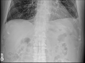
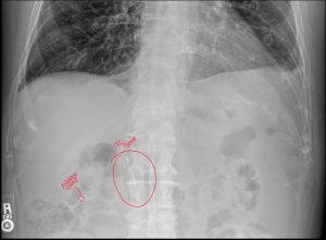
Radiographic analysis identified an incidental inferior vena cava (IVC) filter, an important finding due to its metallic structure that may mimic calcified densities, clips, or vascular changes on plain films. Recognition of this incidental IVC filter prevents misinterpretation, ensuring accurate distinction between foreign devices and potential pathology such as a renal calculus.
This presentation highlights how kidney stones often mimic low back or flank pain, prompting consultations with chiropractors or family physicians before urologists. The case underscores the importance of radiographic interpretation by a board-certified chiropractic radiologist (DACBR) when visceral pathology presents as somatic discomfort.
The leading diagnosis was nephrolithiasis (kidney stone) localized to the right ureter. Differential considerations included:
Using KUB X-ray and diagnostic ultrasound, the DACBR confirmed a radio-opaque calculus adjacent to the right L3 transverse process level—consistent with ureteral positioning. The incidental IVC filter, appearing as a metallic, linear structure near the midline, was duly noted. Its presence reinforced the necessity of careful pattern recognition and contextual imaging review.
For referring chiropractors, deciphering such overlapping findings underscores the value of expert radiology reporting for diagnostic clarity, liability reduction, and interdisciplinary collaboration.
For more on how DACBRs assist healthcare providers in delivering accurate reports, explore About Kinetic Radiology and Resources.
The urinary system includes the kidneys, ureters, bladder, and urethra. Stones develop when mineral crystals, such as calcium oxalate, calcium phosphate, uric acid, or cystine. They aggregate and form hardened deposits within the renal calyces or pelvis.
The formation process involves:
This obstruction triggers hydronephrosis—dilation of the renal pelvis and calyces—and causes the sharp, colicky pain characteristic of renal colic.
In elderly patients, dehydration, reduced renal perfusion, and medication side effects contribute to stone risk. Understanding these mechanisms allows chiropractors and primary care providers to recognize visceral origins of typical “low back pain” presentations.
X-rays remain a frontline diagnostic tool for identifying radio-opaque kidney stones, particularly in chiropractic and outpatient settings.
Common KUB characteristics:
In this case, the right ureteral stone was visible near L3, while the IVC filter projected midline across L2–L3—appearing as metallic filaments consistent with vascular intervention.
DACBRs are adept at differentiating overlapping structures such as:
Radiologists trained in chiropractic radiology provide structured KUB interpretations that clarify findings for clinicians and document incidental findings comprehensively. Learn how our DACBRs approach X-ray interpretation at Kinetic Radiology’s diagnostic imaging page.
Diagnostic ultrasound complements X-ray by visualizing both radiolucent stones and obstructive changes without radiation exposure.
Key ultrasound features in renal calculi:
In this patient, ultrasound revealed a 5 mm hyperechoic shadowing focus in the proximal right ureter and mild hydronephrosis, confirming obstruction. The IVC filter was visible as bright linear echoes within the vena cava, confirming incidental yet benign status.
For chiropractors or family physicians using diagnostic ultrasound services via DACBR teleradiology, these findings empower swift differentiation between renal, vascular, and musculoskeletal causes of back pain.
Explore more about ultrasound and multidisciplinary collaboration in radiology via Resources.
Treatment depends on stone size and location:
Meanwhile, conservative measures like pain management and hydration remain first steps for non-obstructive or mild cases.
Chiropractors and general physicians must:
A DACBR provides a crucial service beyond simply spotting the anomaly. The radiologist’s report will characterize the cervical rib’s morphology (e.g., complete vs. incomplete, presence of a pseudoarthrosis), which has clinical implications. By identifying this key anatomical variant, the DACBR provides the treating clinician with a definitive underlying cause for the patient’s complex symptoms. This diagnostic clarity allows the provider to move beyond non-specific neck and arm pain diagnoses and implement a highly targeted treatment plan addressing the specific biomechanics of the thoracic outlet.
Every day, chiropractors face the same frustration: imaging reports that miss what matters. General radiologists weren’t trained in your world; they don’t understand subluxations, joint dysfunction, or the biomechanical findings that drive your treatment decisions.
The result? Delayed care. Uncertain patients. Cases that stall when they should be progressing.
The Kinetic Radiology Difference: Chiropractors Reading for Chiropractors
Our board-certified DACBRs aren’t just radiologists. We’re chiropractors who chose to specialize in musculoskeletal imaging. We speak your language because we’ve stood where you stand.
Reports You Can Act On Immediately – No vague findings. No irrelevant details. Just the specific insights that guide your next adjustment, your treatment plan, and your patient conversations.
Same-Day Turnaround – Your patients don’t want to wait days wondering what’s wrong. Neither should you. Get clarity fast so care never stalls.
Documentation That Protects Your Practice – Whether it’s insurance requirements, legal protection, or patient records, our reports give you the clinical backing you need.
Confidence That Builds Your Reputation – When patients see you consulting with specialized radiologists, they recognize your commitment to excellence. That trust turns into loyalty, referrals, and five-star reviews.
Think about the last complex case you handled. Did the radiology report actually help you—or did you have to fill in the gaps yourself?
Now imagine having a DACBR partner who catches the subtle findings, flags the red flags, and gives you confidence in every diagnosis.
No commitment. No risk. Just submit your next challenging case and experience what specialized chiropractic radiology can do for your clinical confidence and patient outcomes.
Questions? Call us at 321 325 0096 or email at support@kineticradiology.com
Mineral buildup from dehydration or diet causes stone formation.
Kidney stones occur when urine becomes concentrated with minerals such as calcium, phosphate, or uric acid. In elderly adults, reduced fluid intake, decreased renal function, and medications like diuretics contribute significantly. Age-related metabolic changes also slow urinary flow, creating fertile ground for crystal formation. Identifying these risk factors is crucial, especially when back pain presents atypically in older males and imaging reveals stones incidentally during other evaluations.
Sharp flank or low back pain, hematuria, and urinary urgency are the classic symptoms.
The hallmark symptom of kidney stones is severe, sharp pain that starts in the flank and radiates to the lower abdomen or groin. This pain develops suddenly as the stone moves through the urinary tract, causing spasms and obstruction. Many patients report nausea, vomiting, or visible blood in the urine (hematuria). Because these symptoms can mimic musculoskeletal conditions, they often present first to chiropractors or family doctors. Chiropractors should be alert for cases where “back pain” is not affected by posture or movement and may need imaging evaluation via KUB X-ray, diagnostic ultrasound, or consultation with a DACBR to confirm or rule out a visceral cause.
Kidney stone pain is visceral and non-mechanical, while musculoskeletal pain is movement-dependent.
Musculoskeletal back pain typically worsens with movement or palpation, whereas renal colic is characteristically constant and unrelieved by rest or position changes. Radiating flank pain that follows the dermatomal path but lacks mechanical reproduction should raise suspicion of visceral origin. Using imaging such as a KUB X-ray or ultrasound, a DACBR can help confirm whether the pain is due to a renal calculus, vascular calcification, or lumbar degenerative changes. Chiropractors and family physicians should always include kidney stones in their differential diagnosis for elderly male patients with persistent lower back pain.
A DACBR is a board-certified chiropractic radiologist specializing in imaging interpretation.
A Diplomate of the American Chiropractic Board of Radiology (DACBR) is a chiropractic physician who has completed a three-year full-time postdoctoral residency in diagnostic imaging and passed rigorous national board exams. These professionals interpret X-rays, MRI, CT, CBCT, and diagnostic ultrasound, with a special focus on musculoskeletal and spinal imaging. Importantly, DACBRs also identify incidental findings—like the IVC filter seen in this kidney stone case—ensuring comprehensive reporting that meets both clinical and legal standards. They provide teleradiology reports for chiropractors, physical therapists, and integrative physicians to improve diagnostic accuracy and interprofessional care.
CT is most sensitive, but X-ray and ultrasound remain first-line in most practices.
Although non-contrast CT scans have the highest accuracy for kidney stones, they are not always necessary as an initial test. KUB X-ray and ultrasound are widely used in chiropractic and family medicine for screening and monitoring. X-rays identify most radio-opaque stones, while ultrasound detects both radiolucent stones (like uric acid) and obstruction (hydronephrosis). A DACBR can help interpret these images accurately, providing detailed descriptions that distinguish renal from vascular or skeletal calcifications.
Partnering with a DACBR teleradiology service provides more than just a second opinion; it offers a significant return on investment:
Speed: Get expert reports in hours, not days.
Expertise: Access board-certified specialists without having to hire them.
Convenience: The entire process is handled online from your office.
Clarity: Receive clear, concise reports that are clinically relevant to chiropractic care, not generic medical reports.
Posted onTrustindex verifies that the original source of the review is Google. Kinetic radiology has been an absolute game changer in speed of reports and detailed reports. Any other doctors I send my reports to are amazed at the detail and the pathology that gets picked up. This is my one and only radiologist group, im thrilled.Posted onTrustindex verifies that the original source of the review is Google. Rishi provides an outstanding service—fast, reliable, and incredibly reassuring. He’s quick to respond, efficient in his work, and always takes the time to address any concerns with clarity and professionalism. I highly recommend his services to anyone looking for a dependable DACBR.Posted onTrustindex verifies that the original source of the review is Google. Prompt efficient service that is thorough and clear. Spinal information is top notch and I've had patients discover kidney stones and possible issues with a hip joint replacement loosening as incidental findings that supported both me and the patient above expectations.Posted onTrustindex verifies that the original source of the review is Google. Quick, accurate, and easy to work with. My new radiology team!Posted onTrustindex verifies that the original source of the review is Google. Excellent, timely reads. Invaluable for CBCTPosted onTrustindex verifies that the original source of the review is Google. Best turnaround time and thorough reports out of any radiologist I’ve seen or worked with!Posted onTrustindex verifies that the original source of the review is Google. Very detailed reports and quick service. Highly recommendedPosted onTrustindex verifies that the original source of the review is Google. Fast turn around time for the radiology reports! Thank you for making this process as seamless as possible!Posted onTrustindex verifies that the original source of the review is Google. I am a NUCCA chiropractor located in Wauankee Wisconsin and I can tell you Dr. Rishi is the only radiologist I’d work with. Sure there are many others in my area but when you want the best you go to the best. He is very easy to work with and always fast to respond and report. 100% recommend.Posted onTrustindex verifies that the original source of the review is Google. Kinetic Radiology is great! They were able to read and get a report written immediately. They are my go to company for any and all images that I need read!Load more
We service all 50 U.S. States, including the following States and Cities listed below.
Copyright 2024 Kinetic Radiology All Rights Reserved
Website Privacy | Terms of UseReceive timely resources to keep you and your practice on the cutting edge of Chiropractic Radiology.
Copyright 2024 Kinetic Radiology
All Rights Reserved
Receive timely resources to keep you and your practice on the cutting edge of Chiropractic Radiology.