A 42-year-old female graphic designer presents to your clinic with a primary complaint of debilitating hand pain, which she states began insidiously about 18 months ago but has become acutely inflammatory in the past six months. She describes the pain as a “deep, burning, and gnawing ache” that is most severe in the small joints of her fingers. She reports significant morning stiffness lasting 45-60 minutes, which is accompanied by visible swelling and redness. Functionally, her career is impacted; she struggles with the fine motor control required for using a mouse and stylus and finds it painful to type for extended periods. Activities of daily living, like opening jars or buttoning a shirt, have become difficult.
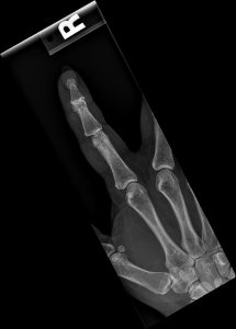
Based on the classic clinical presentation and pathognomonic radiographic findings, the diagnosis is Erosive Osteoarthritis. EOA is a distinct clinical entity, recognized as a subset of osteoarthritis characterized by significant synovial inflammation, leading to aggressive articular cartilage destruction and subchondral bone erosion.
For the chiropractor, who often serves as a primary portal of entry for patients with joint pain, distinguishing EOA from its more common degenerative counterpart and its systemic inflammatory mimics is a critical diagnostic challenge. Mischaracterizing this condition can lead to delayed medical co-management and suboptimal patient outcomes.
This article serves as an in-depth clinical and radiological reference, written from the perspective of a board-certified chiropractic radiologist (DACBR). We will dissect the pathophysiology, clinical presentation, and pathognomonic imaging findings of EOA, providing the detailed knowledge necessary for confident diagnosis and effective interprofessional collaboration.
To understand EOA, one must appreciate that it is a hybrid disease, exhibiting features of both inflammation and degeneration. The process is believed to be initiated by a profound, localized synovitis.
Inflammatory Cascade: While the exact trigger is unknown, the synovial membrane becomes intensely inflamed. This process involves an influx of inflammatory cells and the upregulation of pro-inflammatory cytokines such as Interleukin-1 (IL-1) and Tumor Necrosis Factor-alpha (TNF-α).
Pannus Formation: This chronic synovitis leads to the formation of an aggressive, invasive synovial tissue known as pannus. This tissue is rich in proteolytic enzymes like collagenases and metalloproteinases.
Central Articular Attack: In EOA, the pannus spreads across the articular cartilage surface. It preferentially attacks and erodes the central subchondral bone plate. This is the critical distinction from Rheumatoid Arthritis, where the pannus primarily invades the unprotected “bare areas” at the joint margins.
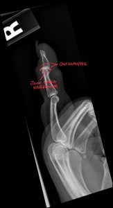
Degenerative Response: Simultaneously, the joint attempts to respond to the instability and damage with classic osteoarthritic changes. This includes subchondral sclerosis (thickening of the bone beneath the cartilage) and the formation of osteophytes (bone spurs) at the joint margins.
This unique combination of central erosive destruction and peripheral productive repair is what creates the hallmark “gull wing” deformity. In the end stage, the joint can undergo complete bony fusion, known as ankylosis.
The radiographic diagnosis of EOA is highly specific. A systematic evaluation of the distribution, erosions, bone density, and joint space is essential.
Distribution: EOA has a strong predilection for the interphalangeal joints of the hands. The DIP and PIP joints are the primary targets. The first carpometacarpal (CMC) joint is also commonly involved. Critically, there is typically sparing of the MCP and wrist joints. This distribution is a key differentiator from RA.
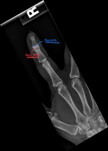
Bone Erosions: The erosions are characteristically central and subchondral. They may initially appear as small pits or irregularities in the central articular surface before coalescing into larger defects.
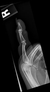
“Gull Wing” Sign: This is the most specific sign of EOA. It represents an advanced stage of central erosion. The proximal phalanx develops a pointed central beak with flared marginal osteophytes, resembling a seagull in flight. The opposing distal phalangeal base develops a corresponding V-shaped central depression.
Bone Density: Bone density is typically normal. The absence of periarticular osteoporosis is a powerful sign that helps exclude RA, where bone demineralization adjacent to inflamed joints is a classic feature.
Cartilage Space: As with other forms of arthritis, there is progressive and often uniform joint space narrowing due to the destruction of articular cartilage.
Soft Tissues: During active inflammatory phases, significant fusiform soft tissue swelling is evident around the affected joints.
While plain radiographs are often diagnostic, advanced imaging can be critical in early disease or atypical presentations.
Magnetic Resonance Imaging (MRI): MRI is the most sensitive modality for detecting early inflammatory changes. It can identify synovitis, joint effusions, and bone marrow edema long before erosions are visible on X-ray. Post-contrast T1-weighted sequences will show intense synovial enhancement, confirming active inflammation.
Diagnostic Ultrasound: High-resolution ultrasound is an excellent, cost-effective tool for evaluating the small joints of the hand. Using Power Doppler, a clinician can visualize and quantify the degree of active synovitis by detecting increased blood flow. Ultrasound can also clearly depict cortical erosions, osteophytes, and tenosynovitis.
Computed Tomography (CT): CT provides unparalleled visualization of bony anatomy. It is superior to X-ray in defining the precise size, shape, and location of central erosions and can be useful in surgical planning for late-stage disease.
| Feature | Erosive Osteoarthritis (EOA) | Rheumatoid Arthritis (RA) | Psoriatic Arthritis (PsA) |
| Primary Distribution | DIPs, PIPs, 1st CMC | MCPs, PIPs, Wrists | Asymmetric; DIPs, “Ray” pattern, Spondylitis |
| Symmetry | Generally Symmetrical | Symmetrical | Asymmetrical |
| Erosion Type | Central, “Gull Wing” | Marginal, “Bare Area” | Marginal, “Pencil-in-Cup,” Aggressive |
| Bone Density | Normal | Periarticular Osteoporosis | Normal; Can have periostitis (“fluffy”) |
| New Bone Formation | Osteophytes (prominent) | Minimal to none | Periostitis (common) |
| Serology | RF/anti-CCP Negative | RF/anti-CCP Often Positive | RF Negative; HLA-B27 may be positive |
| Clinical Clues | Middle-aged women, inflammatory nodes | Systemic symptoms, boggy synovitis | Psoriasis skin/nail lesions, dactylitis |
The management of EOA is multi-faceted and requires a collaborative approach.
Pharmacologic Management: This is the domain of the patient’s primary care physician and rheumatologist. Treatment often begins with NSAIDs. For persistent, aggressive disease, Disease-Modifying Antirheumatic Drugs (DMARDs) such as methotrexate or hydroxychloroquine may be prescribed to control the underlying inflammatory process.
Chiropractic & Physical Medicine: The chiropractor’s role is crucial for managing pain, maintaining function, and addressing secondary biomechanical consequences.
Contraindications: High-velocity, low-amplitude manipulation of the acutely inflamed and eroded small joints of the hand is contraindicated.
Appropriate Care: Management focuses on pain modulation using modalities like low-level laser therapy (photobiomodulation), addressing surrounding soft tissue restrictions in the hand and forearm, and providing gentle mobilization to adjacent non-inflamed joints (wrist, elbow). Addressing compensatory postural changes in the cervical and thoracic spine is also vital.
Referral and Co-Management: Upon forming a diagnosis of EOA, a prompt referral to a rheumatologist is the standard of care. The chiropractor’s role is to make the initial diagnosis, educate the patient about the condition, and facilitate this referral. A detailed radiology report and clinical letter are essential for effective communication. Co-management allows the rheumatologist to control the systemic inflammation while the chiropractor continues to manage the biomechanical and functional aspects of the patient’s care.
In today’s healthcare landscape, collaboration and specialization are key. By integrating Kinetic Radiology into your practice workflow, you gain a powerful partner that enhances your capabilities and builds your reputation.
Unmatched Expertise: Our reports are generated by board-certified chiropractic radiologists (DACBRs). We are chiropractors who have undergone extensive, multi-year residency training focused solely on musculoskeletal diagnostic imaging. We understand the clinical questions you have because we speak your language.
Speed and Certainty: Don’t wait days for a report. Our streamlined process delivers fast, accurate, and actionable Xray reports, allowing you to make confident clinical decisions in real-time.
A Seamless Extension of Your Practice: Think of us as your in-house radiology department. We provide the expertise you need, when you need it, allowing you to focus on what you do best: treating patients. By ordering an expert report, you demonstrate to your patients that you are committed to the highest standard of care.
For your next complex wrist injury, second opinion, or routine Xray report, choose the experts. Partner with Kinetic Radiology. Contact our team of Diagnostic Imaging Consultants today to see how our DACBR-level insights can benefit your patients and your practice.
The exact cause is unknown, but it involves a significant inflammatory component combined with the wear-and-tear of regular OA.
The specific cause of erosive osteoarthritis (EOA) remains idiopathic, meaning it is not fully understood. However, it is recognized as a distinct disease process that combines features of classic degenerative osteoarthritis with a much more aggressive inflammatory component. The primary driver is believed to be a severe, localized synovitis, or inflammation of the joint’s synovial lining. This inflammation is much more intense than in regular OA and leads to the production of enzymes that actively erode the central part of the bone’s articular surface. While not a systemic autoimmune disease like rheumatoid arthritis, risk factors for EOA include being female and postmenopausal, suggesting a hormonal link. A genetic predisposition is also suspected. Understanding that EOA has both degenerative and inflammatory causes is key to its diagnosis and management.
It’s not “worse,” just different; RA is a systemic autoimmune disease, while EOA is a localized, aggressive joint disease.
Comparing the severity of erosive osteoarthritis (EOA) and rheumatoid arthritis (RA) depends on the metric. RA is a systemic autoimmune disease, meaning it can affect not only the joints but also organs like the lungs, heart, and eyes, and it often carries significant systemic symptoms and requires aggressive immunosuppressive medication. In that sense, RA is a more serious systemic condition. However, from a joint-specific perspective, EOA can be extremely destructive and painful. It often leads to severe deformity, bony fusion (ankylosis), and significant functional loss in the hands, which can be just as debilitating as the effects of RA in those joints. The key difference is that EOA’s impact is localized to the joints (primarily the hands), whereas RA is a body-wide disease. A radiology report is crucial for differentiating the two, as their imaging findings are distinct.
Erosive OA has an aggressive inflammatory component that causes central bone erosions, which are completely absent in regular OA.
The primary difference between erosive osteoarthritis (EOA) and standard, non-erosive osteoarthritis (OA) lies in the presence of aggressive inflammation and bone erosion. Regular OA is primarily a degenerative, “wear-and-tear” condition characterized by cartilage loss, joint space narrowing, and the formation of bone spurs (osteophytes). While it can have minor inflammation, it does not destroy the bone itself. EOA, however, features a significant, destructive inflammatory synovitis. This inflammation leads to the hallmark radiographic finding of EOA: central subchondral bone erosions. This process can eventually create the pathognomonic “gull wing” deformity seen on X-rays. Clinically, patients with EOA often experience a more abrupt onset with more intense signs of inflammation, such as redness, warmth, and severe pain, compared to the slow, progressive stiffness typical of regular OA.
Modern teleradiology platforms make getting an expert second opinion simple, fast, and secure.
Find a Chiropractic Teleradiology Service: Choose a reputable service that is staffed by DACBRs. These platforms are designed specifically for chiropractors.
Securely Upload Patient Images: Export the DICOM files from your imaging software and upload them to the service’s secure, HIPAA-compliant online portal. You will also provide a brief clinical history for context.
Receive Your Detailed Report: The DACBR interprets the images and sends a comprehensive, actionable report directly to you, often within 24 hours. This report will clearly state the findings, impressions, and clinical recommendations.
Partnering with a DACBR teleradiology service provides more than just a second opinion; it offers a significant return on investment:
Speed: Get expert reports in hours, not days.
Expertise: Access board-certified specialists without having to hire them.
Convenience: The entire process is handled online from your office.
Clarity: Receive clear, concise reports that are clinically relevant to chiropractic care, not generic medical reports.
Posted onTrustindex verifies that the original source of the review is Google. Kinetic radiology has been an absolute game changer in speed of reports and detailed reports. Any other doctors I send my reports to are amazed at the detail and the pathology that gets picked up. This is my one and only radiologist group, im thrilled.Posted onTrustindex verifies that the original source of the review is Google. Rishi provides an outstanding service—fast, reliable, and incredibly reassuring. He’s quick to respond, efficient in his work, and always takes the time to address any concerns with clarity and professionalism. I highly recommend his services to anyone looking for a dependable DACBR.Posted onTrustindex verifies that the original source of the review is Google. Prompt efficient service that is thorough and clear. Spinal information is top notch and I've had patients discover kidney stones and possible issues with a hip joint replacement loosening as incidental findings that supported both me and the patient above expectations.Posted onTrustindex verifies that the original source of the review is Google. Quick, accurate, and easy to work with. My new radiology team!Posted onTrustindex verifies that the original source of the review is Google. Excellent, timely reads. Invaluable for CBCTPosted onTrustindex verifies that the original source of the review is Google. Best turnaround time and thorough reports out of any radiologist I’ve seen or worked with!Posted onTrustindex verifies that the original source of the review is Google. Very detailed reports and quick service. Highly recommendedPosted onTrustindex verifies that the original source of the review is Google. Fast turn around time for the radiology reports! Thank you for making this process as seamless as possible!Posted onTrustindex verifies that the original source of the review is Google. I am a NUCCA chiropractor located in Wauankee Wisconsin and I can tell you Dr. Rishi is the only radiologist I’d work with. Sure there are many others in my area but when you want the best you go to the best. He is very easy to work with and always fast to respond and report. 100% recommend.Posted onTrustindex verifies that the original source of the review is Google. Kinetic Radiology is great! They were able to read and get a report written immediately. They are my go to company for any and all images that I need read!Load more
We service all 50 U.S. States, including the following States and Cities listed below.
Copyright 2024 Kinetic Radiology All Rights Reserved
Website Privacy | Terms of UseReceive timely resources to keep you and your practice on the cutting edge of Chiropractic Radiology.
Copyright 2024 Kinetic Radiology
All Rights Reserved
Receive timely resources to keep you and your practice on the cutting edge of Chiropractic Radiology.