A 35-year-old male presents with acute chest and rib pain following a motor vehicle accident. The patient reports difficulty with breathing, with a catching-like pain wrapping around his right ribs. Physical examination reveal mild ecchymosis at the site of pain. Pulmonary exam is unremarkable.
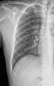
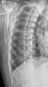
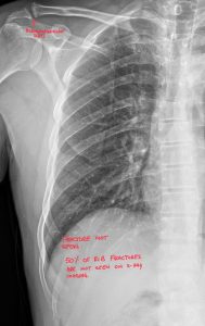
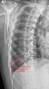
The oblique rib views demonstrate a rib fracture of the right 10th rib with mild cortical offset. The PA rib x-ray does not demonstrate this finding.
Rib fractures are a common musculoskeletal injury we see frequently in Port Orange, Florida, often resulting from falls, car accidents, or even strenuous activity. While the clinical presentation can range from obvious to subtle, accurately diagnosing these fractures through diagnostic imaging is crucial for effective patient management. This guide is tailored for chiropractors and other healthcare professionals in the Port Orange area, focusing on the radiographic nuances of rib fractures, the subtle signs to watch for, associated soft tissue injuries, and the growing role of diagnostic ultrasound, alongside the indispensable expertise of diagnostic imaging consultants.
Our initial imaging approach for suspected rib fractures in Port Orange typically involves standard chest X-rays (PA and lateral views). While these are valuable for identifying displaced fractures, many rib fractures present with remarkably subtle or even hidden findings.
Even when a direct fracture line isn’t immediately apparent, being aware of indirect signs on chest X-ray can raise suspicion:
Rib fractures don’t occur in isolation. Associated soft tissue changes can be critical clues, especially when bony findings are subtle on radiographs.
While X-rays are the first line, CT scans offer superior detail for detecting rib fractures and associated injuries, especially in:
Suspected Pathological Fractures: To better assess the underlying bone structure.
Diagnostic ultrasound is increasingly being recognized as a valuable tool for evaluating rib fractures, particularly in specific clinical scenarios. It offers several advantages:
It’s important to acknowledge the limitations of ultrasound:
Ultrasound can be a useful adjunct to radiography, particularly in:
Follow-up of Known Fractures: To assess for healing and potential complications like hematoma formation.
In a diverse healthcare setting, collaborating with a diagnostic imaging consultant, such as a DACBR chiropractic radiologist with expertise in musculoskeletal imaging, offers significant benefits:
Improved Communication and Clinical Correlation: Radiologists can provide crucial context, linking imaging findings to the patient’s clinical presentation
In today’s healthcare landscape, collaboration and specialization are key. By integrating Kinetic Radiology into your practice workflow, you gain a powerful partner that enhances your capabilities and builds your reputation.
Unmatched Expertise: Our reports are generated by board-certified chiropractic radiologists (DACBRs). We are chiropractors who have undergone extensive, multi-year residency training focused solely on musculoskeletal diagnostic imaging. We understand the clinical questions you have because we speak your language.
Speed and Certainty: Don’t wait days for a report. Our streamlined process delivers fast, accurate, and actionable Xray reports, allowing you to make confident clinical decisions in real-time.
A Seamless Extension of Your Practice: Think of us as your in-house radiology department. We provide the expertise you need, when you need it, allowing you to focus on what you do best: treating patients. By ordering an expert report, you demonstrate to your patients that you are committed to the highest standard of care.
For your next complex wrist injury, second opinion, or routine Xray report, choose the experts. Partner with Kinetic Radiology. Contact our team of Diagnostic Imaging Consultants today to see how our DACBR-level insights can benefit your patients and your practice.
Rib fractures are hard to see on X-rays due to overlapping bones and soft tissues, subtle or non-displaced fractures, and the orientation of the fracture line, all of which can obscure the injury.
The main symptom is sharp, localized chest or rib pain that worsens with deep breathing, coughing, or movement. Patients may also experience tenderness, bruising, or a catching-like pain.
Yes, it’s very common to have a rib fracture that doesn’t appear on a standard X-ray. When this happens, advanced diagnostic imaging like a CT scan or ultrasound is crucial to confirm the injury and ensure proper management.
Modern teleradiology platforms make getting an expert second opinion simple, fast, and secure.
Find a Chiropractic Teleradiology Service: Choose a reputable service that is staffed by DACBRs. These platforms are designed specifically for chiropractors.
Securely Upload Patient Images: Export the DICOM files from your imaging software and upload them to the service’s secure, HIPAA-compliant online portal. You will also provide a brief clinical history for context.
Receive Your Detailed Report: The DACBR interprets the images and sends a comprehensive, actionable report directly to you, often within 24 hours. This report will clearly state the findings, impressions, and clinical recommendations.
Partnering with a DACBR teleradiology service provides more than just a second opinion; it offers a significant return on investment:
Speed: Get expert reports in hours, not days.
Expertise: Access board-certified specialists without having to hire them.
Convenience: The entire process is handled online from your office.
Clarity: Receive clear, concise reports that are clinically relevant to chiropractic care, not generic medical reports.
Posted onTrustindex verifies that the original source of the review is Google. Kinetic radiology has been an absolute game changer in speed of reports and detailed reports. Any other doctors I send my reports to are amazed at the detail and the pathology that gets picked up. This is my one and only radiologist group, im thrilled.Posted onTrustindex verifies that the original source of the review is Google. Rishi provides an outstanding service—fast, reliable, and incredibly reassuring. He’s quick to respond, efficient in his work, and always takes the time to address any concerns with clarity and professionalism. I highly recommend his services to anyone looking for a dependable DACBR.Posted onTrustindex verifies that the original source of the review is Google. Prompt efficient service that is thorough and clear. Spinal information is top notch and I've had patients discover kidney stones and possible issues with a hip joint replacement loosening as incidental findings that supported both me and the patient above expectations.Posted onTrustindex verifies that the original source of the review is Google. Quick, accurate, and easy to work with. My new radiology team!Posted onTrustindex verifies that the original source of the review is Google. Excellent, timely reads. Invaluable for CBCTPosted onTrustindex verifies that the original source of the review is Google. Best turnaround time and thorough reports out of any radiologist I’ve seen or worked with!Posted onTrustindex verifies that the original source of the review is Google. Very detailed reports and quick service. Highly recommendedPosted onTrustindex verifies that the original source of the review is Google. Fast turn around time for the radiology reports! Thank you for making this process as seamless as possible!Posted onTrustindex verifies that the original source of the review is Google. I am a NUCCA chiropractor located in Wauankee Wisconsin and I can tell you Dr. Rishi is the only radiologist I’d work with. Sure there are many others in my area but when you want the best you go to the best. He is very easy to work with and always fast to respond and report. 100% recommend.Posted onTrustindex verifies that the original source of the review is Google. Kinetic Radiology is great! They were able to read and get a report written immediately. They are my go to company for any and all images that I need read!Load more
We service all 50 U.S. States, including the following States and Cities listed below.
Copyright 2024 Kinetic Radiology All Rights Reserved
Website Privacy | Terms of UseReceive timely resources to keep you and your practice on the cutting edge of Chiropractic Radiology.
Copyright 2024 Kinetic Radiology
All Rights Reserved
Receive timely resources to keep you and your practice on the cutting edge of Chiropractic Radiology.