A 29-year-old male warehouse worker presents with acute right lateral foot and ankle pain. He states he was stepping down from a loading dock, missed the last step, and forcefully “rolled” his ankle inwards. He reports immediate, sharp pain and difficulty bearing weight. His initial evaluation at an urgent care center the day prior resulted in a diagnosis of a “severe ankle sprain,” with instructions for rest and ice. Standard AP, lateral, and mortise ankle radiographs were performed at that time. He presents to your office for a second opinion and to initiate a workers’ compensation claim.
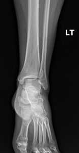
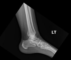

Acute avulsion fracture at the dorsal calcaneocuboid joint, consistent with a tear of the calcaneocuboid ligament.
In the context of personal injury and workers’ compensation, the accurate and timely diagnosis of a bony injury is paramount. While ligamentous sprains are common, they are often considered subjective injuries, leading to disputes over causation and disability. The cuboid avulsion fracture, however, represents an objective, undeniable sign of significant trauma. It is one of the most frequently missed fractures in the foot, as its symptoms masterfully mimic a simple lateral ankle sprain.¹
The failure to identify this fracture can lead to chronic lateral foot pain, nonunion of the fragment, and significant functional impairment for the patient. For the treating clinician, a missed diagnosis can result in a weakened medicolegal case for the patient, potentially depriving them of appropriate compensation and care. A high index of suspicion and a commitment to obtaining the correct imaging views are therefore essential. This clinic will review the anatomy, clinical findings, and critical imaging pearls for identifying this “hidden” fracture, emphasizing its importance in the personal injury and workers’ compensation arena.
The cuboid bone is a critical component of the lateral column of the foot, articulating with the calcaneus proximally and the fourth and fifth metatarsals distally. It acts as a keystone, providing structural stability during gait. The dorsal surface of the cuboid is the attachment site for the dorsal calcaneocuboid ligament, a broad, strong ligament that reinforces the calcaneocuboid joint capsule.
A cuboid avulsion fracture typically occurs via one of two mechanisms:
Forceful Inversion and Plantar-flexion: This is the most common mechanism, identical to a classic ankle sprain. As the foot rolls inward, immense tension is placed on the dorsal calcaneocuboid ligament. If the force exceeds the shear strength of the bone, the ligament pulls a fragment of bone off the dorsal cuboid or the anterior process of the calcaneus.²
“Nutcracker” Injury: This occurs from forced abduction of the forefoot, which compresses the cuboid between the calcaneus and the metatarsals. This compressive force can cause the cuboid to fracture.
The clinical presentation is what makes this fracture so deceptive. Patients almost universally report a history of an “ankle roll” or twisting injury. The signs and symptoms overlap almost perfectly with a lateral ankle sprain:
Localized pain on the lateral aspect of the midfoot, just distal to the lateral malleolus.
Swelling and ecchymosis (bruising) in the same area.
Point tenderness directly over the calcaneocuboid joint.
Difficulty and pain with weight-bearing.
Because these findings are so common, many clinicians may prematurely conclude the diagnosis is a sprain without investigating further. The key to diagnosis is maintaining a high index of suspicion in any patient with lateral foot pain after an inversion injury, especially when the point of maximal tenderness is over the midfoot rather than directly over the ankle ligaments.
Proper imaging is the cornerstone of diagnosis. As demonstrated in the clinical history, a standard ankle radiographic series is often insufficient and can be misleadingly negative.
Ankle Series (AP, Lateral, Mortise): These views are designed to evaluate the tibiotalar and fibulotalar joints. They often fail to profile the calcaneocuboid joint properly, and a small avulsion fragment can be easily obscured by overlying bony structures.
Foot Series (AP, Oblique, Lateral): A dedicated foot series is essential. The single most important view for diagnosing a cuboid avulsion fracture is the medial oblique view. This projection rotates the foot to provide a clear, unobscured profile of the calcaneocuboid joint. The clinician should carefully inspect the dorsal margin of this joint for a small, osseous fleck.
Differential Diagnosis on Imaging: It is important to differentiate an acute avulsion from normal anatomical variants, such as an os peroneum, which is an accessory ossicle found within the peroneus longus tendon. An os peroneum is typically well-corticated with smooth, rounded edges, whereas an acute fracture fragment will have sharp, irregular margins.
The management of a cuboid avulsion fracture must address both the clinical injury and its significant medicolegal context.
Clinical Treatment: Most cuboid avulsion fractures are treated conservatively. Management includes a period of immobilization in a walking boot or short leg cast for 4 to 6 weeks. An initial period of non-weight-bearing is often required to minimize pain and allow for healing. Physical therapy is initiated after immobilization to restore range of motion, strength, and proprioception.
Personal Injury & Workers’ Compensation Implications: The diagnosis of a fracture, rather than a sprain, fundamentally changes the case.
Objective Proof: The fracture serves as objective, irrefutable evidence of a significant injury directly linked to the traumatic event.
Disability and Work Status: A sprain might be managed with an ankle brace and immediate return to modified duty. A fracture requires a definitive period of immobilization and non-weight-bearing, justifying a longer period of temporary disability and clear work restrictions.
Impairment Rating: A documented fracture will result in a higher permanent impairment rating than a ligamentous sprain according to most jurisdictional guidelines, which directly impacts the final settlement or award.
Documentation: Your report must clearly state “acute avulsion fracture of the cuboid” and describe the imaging findings. This clarity is essential for insurance adjusters, attorneys, and case managers.
For any chiropractor dedicated to providing the highest level of care, knowing when to seek an expert opinion is as important as knowing how to read an X-ray. This is where the specialized expertise of a DACBR becomes an invaluable asset to your practice.
A DACBR is a Diplomate of the American Chiropractic Board of Radiology. This credential signifies that a Doctor of Chiropractic has completed a rigorous, multi-year, full-time residency in clinical radiology and has passed demanding board examinations. Their training is focused specifically on the application of diagnostic imaging within the chiropractic scope of practice, making them the ultimate specialists in musculoskeletal imaging for the profession.
When you send a case for review, you need more than a generic description. You need a clinically relevant interpretation. A radiology report authored by a DACBR is tailored to the needs of a chiropractor. It will not only describe the findings but also:
Provide a concise, confident diagnosis or a relevant list of differential diagnoses.
Answer the specific clinical question you have about your patient.
Place the findings in the context of chiropractic management.
Recommend further imaging or co-management if necessary.
This detailed report becomes a permanent part of the patient’s record, providing legal protection and enhancing interprofessional communication.
Engaging Diagnostic Imaging Consultants, especially those who are DACBR-certified, is a wise decision in several scenarios:
When findings are subtle or atypical.
When you need to definitively differentiate gout from a close mimic like septic arthritis.
To confirm a diagnosis before initiating a long-term management plan.
For complex cases involving multiple comorbidities.
At KineticRadiology.com, we connect you with these radiology specialists, ensuring you have the diagnostic confidence to manage your patients effectively.
The cuboid avulsion fracture is a significant injury often disguised as a simple ankle sprain. For chiropractors and medical providers managing personal injury and workers’ compensation patients, its identification is critical. Remember these key points:
Maintain a High Index of Suspicion: Any patient with lateral foot pain after an inversion injury, especially with point tenderness over the midfoot, should be evaluated for a cuboid fracture.
Order the Correct Views: A standard ankle series is not enough. A dedicated foot series, including a medial oblique view, is essential for diagnosis.
Recognize the Medicolegal Importance: This diagnosis provides objective proof of injury, justifies specific treatment and work restrictions, and increases the value and validity of a personal injury or workers’ compensation claim.
From a subtle cuboid avulsion fracture disguised as an ankle sprain to a spinal teardrop fracture hidden within a whiplash injury, it is clear that these bony injuries are far more than minor “chips.” They are definitive, objective proof of significant trauma. In the demanding arenas of personal injury and workers’ compensation, identifying these fractures is not just good clinical practice; it is a mandate for providing excellent patient care and thorough medicolegal documentation.
However, making that definitive diagnosis, especially from plain film radiographs, requires a specialized eye trained to see what others might miss. This is where the expertise of a board-certified chiropractic radiologist, a DACBR, becomes your greatest asset.
At Kineticradiology.com, we bridge the gap between your clinical suspicion and diagnostic certainty. Our team of DACBR specialists provides more than just a standard radiology report; we provide clarity. A detailed DACBR report from our consultants will not only confirm the presence of an avulsion fracture but will also describe its precise characteristics in a way that is clear, defensible, and tailored for the medicolegal environment.
Elevate your practice, protect your patients, and strengthen your documentation by partnering with the experts. Visit Kineticradiology.com today to learn how our teleradiology services can provide you with the diagnostic confidence you and your patients deserve.
The first step for a chiropractor is to maintain a high index of suspicion and order a dedicated foot radiographic series, not just an ankle series. It is essential to include a medial oblique view. The second step, due to the subtlety of the injury and the high stakes in a workers’ compensation case, is to obtain an expert radiology interpretation from a specialist like a DACBR.
This is a classic scenario. Standard ankle X-rays are not designed to properly visualize the midfoot. A cuboid avulsion fracture is frequently missed on these views. For any personal injury claim, it is crucial to image the specific area of tenderness. Ordering a new foot series with an oblique view is necessary to rule out a fracture and ensure the full extent of the injury is documented.
A chiropractor plays a crucial co-management role. While direct manipulation of the fractured cuboid is contraindicated during the acute healing phase, the chiropractor can treat associated biomechanical issues in the kinetic chain, such as sacroiliac or lumbar dysfunction resulting from altered gait. This care is often essential for a full recovery in a personal injury patient.
The gold standard process is:
1) Maintain a high index of suspicion based on the mechanism of injury.
2) Order a complete and specific radiographic series for the area of maximal tenderness.
3) Obtain a specialty radiology interpretation from a qualified DACBR to get a definitive diagnosis and a defensible report.
4) Based on the DACBR report, initiate a co-management plan with an orthopedist and document all findings meticulously for the personal injury or workers’ comp case.
Posted onTrustindex verifies that the original source of the review is Google. Kinetic radiology has been an absolute game changer in speed of reports and detailed reports. Any other doctors I send my reports to are amazed at the detail and the pathology that gets picked up. This is my one and only radiologist group, im thrilled.Posted onTrustindex verifies that the original source of the review is Google. Rishi provides an outstanding service—fast, reliable, and incredibly reassuring. He’s quick to respond, efficient in his work, and always takes the time to address any concerns with clarity and professionalism. I highly recommend his services to anyone looking for a dependable DACBR.Posted onTrustindex verifies that the original source of the review is Google. Prompt efficient service that is thorough and clear. Spinal information is top notch and I've had patients discover kidney stones and possible issues with a hip joint replacement loosening as incidental findings that supported both me and the patient above expectations.Posted onTrustindex verifies that the original source of the review is Google. Quick, accurate, and easy to work with. My new radiology team!Posted onTrustindex verifies that the original source of the review is Google. Excellent, timely reads. Invaluable for CBCTPosted onTrustindex verifies that the original source of the review is Google. Best turnaround time and thorough reports out of any radiologist I’ve seen or worked with!Posted onTrustindex verifies that the original source of the review is Google. Very detailed reports and quick service. Highly recommendedPosted onTrustindex verifies that the original source of the review is Google. Fast turn around time for the radiology reports! Thank you for making this process as seamless as possible!Posted onTrustindex verifies that the original source of the review is Google. I am a NUCCA chiropractor located in Wauankee Wisconsin and I can tell you Dr. Rishi is the only radiologist I’d work with. Sure there are many others in my area but when you want the best you go to the best. He is very easy to work with and always fast to respond and report. 100% recommend.Posted onTrustindex verifies that the original source of the review is Google. Kinetic Radiology is great! They were able to read and get a report written immediately. They are my go to company for any and all images that I need read!Load more
We service all 50 U.S. States, including the following States and Cities listed below.
Copyright 2024 Kinetic Radiology All Rights Reserved
Website Privacy | Terms of UseReceive timely resources to keep you and your practice on the cutting edge of Chiropractic Radiology.
Copyright 2024 Kinetic Radiology
All Rights Reserved
Receive timely resources to keep you and your practice on the cutting edge of Chiropractic Radiology.