A 34-year-old male presents with severe right wrist pain following a sports injury.
The patient was playing soccer earlier today and was running at full speed when he was tripped, causing him to fall forward. He extended his right arm to break the fall, impacting the hard ground directly onto his palm. He reports immediate, sharp pain and was unable to continue playing.
On examination, there is significant soft tissue swelling and early bruising around the entire wrist joint. There is no gross deformity, but the patient exhibits exquisite tenderness to palpation directly over the distal radius and the radiocarpal joint line. He is unable to make a fist or bear any weight on the hand, and both active and passive range of motion are severely limited by pain. Distal neurovascular status is intact.
Acute distal radius fracture.
A distal radial fracture is a break in the radius bone, near the wrist. It accounts for an estimated 25% of all fractures in the pediatric population and is the single most common fracture seen in the elderly. The classic mechanism of injury is a fall on an outstretched hand (FOOSH) and is something you undoubtedly hear about in your practice. While some cases present with obvious deformity, many are subtle, masquerading as a simple “wrist sprain.” This is where your role becomes pivotal. An unmanaged or mismanaged fracture can lead to chronic pain, stiffness, deformity, and long-term disability for your patient.
Accurate and timely diagnosis is non-negotiable. While many chiropractors are proficient in taking and interpreting radiographs, complex cases or subtle findings can create diagnostic uncertainty. This is where the specialized expertise of a board-certified chiropractic radiologist, or DACBR, becomes an invaluable asset. Throughout this article, we’ll unpack the details of this common injury and demonstrate how leveraging a service like Kinetic Radiology for your reads and second opinions empowers you to practice at the top of your license.
The incidence of distal radial fractures follows a bimodal distribution. The first peak occurs in younger, active individuals, often resulting from high-energy trauma like sports injuries or motor vehicle accidents. The second, more prominent peak, occurs in the elderly population, particularly postmenopausal women, where the fracture is often a consequence of low-energy trauma superimposed on osteoporotic bone.
For your chiropractic practice, the clinical relevance is twofold:
Direct Presentation: Patients may come to you directly after a fall, believing they have a sprain. Your ability to differentiate between a ligamentous injury and a fracture is crucial for immediate, appropriate triage and referral. Missing a fracture not only delays proper care but also carries significant medicolegal risk.
Post-Injury Rehabilitation: You are uniquely positioned to manage the entire kinetic chain after the fracture has healed and the patient is out of their cast. Addressing compensatory patterns in the elbow, shoulder, and cervical spine is essential for a full functional recovery, making you a key player in the patient’s multidisciplinary care team.
Understanding the nuances of this injury allows you to confidently guide patients, whether that means referring them for orthopedic consultation or developing a comprehensive post-immobilization rehabilitation plan.
A solid grasp of the distal radius anatomy is the foundation for accurate radiographic interpretation. This isn’t just about identifying the bone; it’s about understanding the key articular relationships and structural landmarks that determine stability and function.
Articular Surface: The distal radius has two primary facets that form the roof of the radiocarpal joint: a larger scaphoid facet and a smaller lunate facet. The integrity of this surface is critical; intra-articular fractures (those that disrupt the joint surface) have a higher risk of post-traumatic osteoarthritis.
Radial Styloid: This prominent projection on the lateral aspect of the radius serves as an attachment point for the brachioradialis tendon and the radial collateral ligament, providing key stability to the wrist.
Ulnar Notch: A concave surface on the medial aspect of the distal radius that articulates with the head of the ulna, forming the distal radioulnar joint (DRUJ). The DRUJ is essential for forearm pronation and supination.
Lister’s Tubercle: A small bony prominence on the dorsal surface that acts as a pulley for the extensor pollicis longus tendon.
Radial Inclination (or Radial Angle): On a posteroanterior (PA) view, this is the angle between a line perpendicular to the long axis of the radius and a line drawn between the tips of the radial styloid and the ulnar corner of the lunate facet. The normal angle is approximately 21-25 degrees. A loss of this angle indicates fracture collapse and radial shortening.
Volar Tilt (or Palmar Tilt): On a lateral view, this is the angle between a line perpendicular to the long axis of the radius and a line connecting the dorsal and volar rims of the articular surface. The normal orientation is a volar tilt of about 10-15 degrees. In many FOOSH injuries, this tilt is lost or even reversed into dorsal angulation.
Understanding these normal values is what allows you to recognize the pathological changes present in a fracture.
The story the patient tells often points directly to the diagnosis. The vast majority of distal radial fractures result from a FOOSH. The position of the wrist at the time of impact dictates the resulting fracture pattern.
Extension Injury (FOOSH with wrist in extension): This is the most common mechanism, leading to the classic Colles’ fracture. The force causes the distal fragment to be displaced and angulated dorsally. Patients often present with the characteristic “dinner fork” or “bayonet” deformity, where the wrist has a visible dorsal bump.
Flexion Injury (Fall on the back of the hand): This less common mechanism, with the wrist in flexion, results in a Smith’s fracture. Here, the distal fragment is displaced and angulated volarly (palmar). This is often called a “reverse Colles'” and can present with a “garden spade” deformity.
History of a fall or direct trauma.
Immediate, severe pain at the wrist.
Rapid onset of swelling and ecchymosis.
Visible deformity (though not always present, especially in non-displaced fractures).
Marked tenderness to palpation over the distal radius.
Crepitus with movement.
Inability or significant pain with attempts to move the wrist or hand.
It’s also critical to perform a basic neurovascular assessment, checking for sensation in the fingers and capillary refill, as median nerve compromise can occur.
Diagnostic imaging is not just confirmatory; it’s essential for classification, guiding treatment, and identifying associated injuries.
Plain film radiography is the primary and most important imaging modality for suspected distal radial fractures. A standard series should always include:
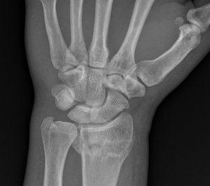
Posteroanterior (PA) View: Excellent for assessing radial inclination, radial shortening, and fracture lines involving the radial styloid or articular surface.
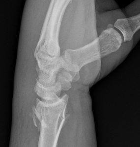
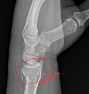
Lateral View: The most critical view for assessing volar tilt and determining dorsal or volar displacement/angulation.
Oblique View: Can help to better visualize specific parts of the articular surface and identify fracture lines that may be obscured on the standard two views.
When you order an X-ray report from Kinetic Radiology, our DACBR team scrutinizes each of these views to provide a comprehensive and clinically relevant interpretation.
While X-ray is the starting point, it doesn’t always tell the whole story.
Computed Tomography (CT): CT is the gold standard for evaluating complex, comminuted (multiple fragments), and intra-articular fractures. It provides exquisite bony detail, allowing an orthopedic surgeon to fully understand the fracture pattern and plan for surgical fixation (ORIF – Open Reduction Internal Fixation).
Magnetic Resonance Imaging (MRI): MRI is indicated when there is a strong clinical suspicion of a significant soft tissue injury that could affect stability or outcomes. This includes tears of the Triangular Fibrocartilage Complex (TFCC), the scapholunate ligament, or the lunotriquetral ligament. It is also the most sensitive modality for identifying occult (hidden) fractures not visible on plain films.
Developing a systematic approach to reviewing wrist X-rays will prevent you from missing key findings. Think of it as a consistent checklist for every film.
A – Alignment: First, assess the overall alignment. Look at the lateral view. Is there normal volar tilt, or is there dorsal angulation? Look at the PA view. Is there a loss of radial inclination? Is the radius shortened relative to the ulna (positive ulnar variance)?
B – Bones: Trace the cortex of the radius and ulna on all views. Look for any break, buckle, or lucent line. Is the fracture extra-articular (sparing the joint) or intra-articular (entering the joint)? Is it a simple two-part fracture or comminuted? Don’t forget to carefully inspect the ulnar styloid—a fracture here is present in over 50% of cases and can imply instability of the DRUJ and TFCC.
C – Cartilage (Joint Spaces): Assess the radiocarpal and distal radioulnar joint spaces. Are they maintained and congruent? Widening of the scapholunate interval (>3 mm) can indicate a ligamentous tear, a significant associated injury.
Colles’ Fracture: The most common type. Characterized by an extra-articular fracture with dorsal displacement and angulation of the distal fragment.
Smith’s Fracture: The “reverse Colles’.” An extra-articular fracture with volar displacement and angulation.
Barton’s Fracture: An intra-articular fracture involving the dorsal or volar rim of the distal radius. It is an unstable fracture-dislocation of the radiocarpal joint.
Chauffeur’s (Hutchinson’s) Fracture: An oblique, intra-articular fracture of the radial styloid.
Recognizing these patterns is important, but describing the fracture based on its anatomical characteristics (intra-articular vs. extra-articular, displacement, angulation, comminution) is more clinically useful.
You’ve taken good quality X-rays, but something doesn’t feel right. Maybe the patient’s pain is far greater than what the films seem to show, or perhaps the fracture pattern is confusing. This is the perfect time to seek the opinion of a DACBR.
High Clinical Suspicion, Negative X-rays: A patient with a clear FOOSH mechanism and exquisite snuffbox or distal radial tenderness but negative initial films. An occult scaphoid or radial fracture is possible, and a DACBR can recommend the next best step, whether it’s immobilization and repeat films in 10-14 days or proceeding to MRI.
Complex Intra-articular Fractures: When the joint surface is clearly involved and there are multiple fragments, a detailed description is crucial for the receiving orthopedist. The Diagnostic Imaging Consultants at Kinetic Radiology can provide a report that precisely details the extent of articular involvement.
Suspected Ligamentous Injury: If you notice subtle signs of instability on the X-ray, like a widened scapholunate space, a DACBR can confirm your suspicions and highlight the need for further evaluation, often with MRI.
Medicolegal Peace of Mind: For any fracture, having a definitive X-ray report from a board-certified radiologist in your patient’s file is the standard of care and your best protection. It demonstrates diligence and ensures your clinical decisions are based on an expert interpretation.
Partnering with Kinetic Radiology means you have a DACBR on your team, ready to provide the clarity and confidence you need to manage these cases effectively.
Your role as the chiropractor is not to set the fracture, but to be an expert diagnostician and coordinator of care.
Initial Diagnosis: Obtain high-quality radiographs and make a provisional diagnosis or, ideally, get an immediate STAT Xray report from a service like Kinetic Radiology.
Immobilization and Referral: Place the patient in a temporary splint to provide comfort and prevent further injury. Based on the fracture severity, refer them to the appropriate setting—an urgent care center for a simple, non-displaced fracture that may only need casting, or the emergency department for a significantly displaced or unstable fracture.
Collaboration: Communicate your findings clearly to the receiving provider. A detailed report from one of our Diagnostic Imaging Consultants is the perfect tool for this, ensuring a seamless handoff.
Post-Immobilization Care: This is where you shine. Once the orthopedist clears the patient for rehabilitation (typically 6-8 weeks post-injury), you can implement a protocol to:
Restore wrist and finger range of motion.
Improve grip strength.
Address secondary biomechanical issues in the elbow, shoulder, and cervical/thoracic spine that developed as a result of immobilization and compensation.
In today’s healthcare landscape, collaboration and specialization are key. By integrating Kinetic Radiology into your practice workflow, you gain a powerful partner that enhances your capabilities and builds your reputation.
Unmatched Expertise: Our reports are generated by board-certified chiropractic radiologists (DACBRs). We are chiropractors who have undergone extensive, multi-year residency training focused solely on musculoskeletal diagnostic imaging. We understand the clinical questions you have because we speak your language.
Speed and Certainty: Don’t wait days for a report. Our streamlined process delivers fast, accurate, and actionable Xray reports, allowing you to make confident clinical decisions in real-time.
A Seamless Extension of Your Practice: Think of us as your in-house radiology department. We provide the expertise you need, when you need it, allowing you to focus on what you do best: treating patients. By ordering an expert report, you demonstrate to your patients that you are committed to the highest standard of care.
For your next complex wrist injury, second opinion, or routine Xray report, choose the experts. Partner with Kinetic Radiology. Contact our team of Diagnostic Imaging Consultants today to see how our DACBR-level insights can benefit your patients and your practice.
While both can be painful, signs of a fracture are often more severe. Look for:
Intense pain immediately after an impact or fall.
Significant swelling that appears quickly.
A “crack” or “snap” sound at the time of injury.
A visible deformity, where your wrist looks crooked or bent.
An inability to move or put any weight on your wrist.
Ultimately, the only way to know for sure is with an X-ray. If you suspect a fracture, it’s critical to see a healthcare provider right away for an accurate diagnosis.
The bone itself typically takes about 6 to 8 weeks to heal, and you’ll likely be in a cast or splint during this time. However, total recovery takes longer. Regaining your full range of motion and strength can take several months of dedicated rehabilitation after the cast is removed.
Once your orthopedic doctor has confirmed the bone is healed and has removed your cast, a chiropractor can be a key part of your recovery team. We focus on:
Restoring range of motion with gentle joint mobilization for the wrist, hand, and elbow.
Performing soft tissue therapy to break down scar tissue.
Prescribing specific exercises to rebuild grip strength and stability.
Addressing compensatory pain that may have developed in your elbow, shoulder, or neck from being in a sling.
Modern teleradiology platforms make getting an expert second opinion simple, fast, and secure.
Find a Chiropractic Teleradiology Service: Choose a reputable service that is staffed by DACBRs. These platforms are designed specifically for chiropractors.
Securely Upload Patient Images: Export the DICOM files from your imaging software and upload them to the service’s secure, HIPAA-compliant online portal. You will also provide a brief clinical history for context.
Receive Your Detailed Report: The DACBR interprets the images and sends a comprehensive, actionable report directly to you, often within 24 hours. This report will clearly state the findings, impressions, and clinical recommendations.
Partnering with a DACBR teleradiology service provides more than just a second opinion; it offers a significant return on investment:
Speed: Get expert reports in hours, not days.
Expertise: Access board-certified specialists without having to hire them.
Convenience: The entire process is handled online from your office.
Clarity: Receive clear, concise reports that are clinically relevant to chiropractic care, not generic medical reports.
Posted onTrustindex verifies that the original source of the review is Google. Kinetic radiology has been an absolute game changer in speed of reports and detailed reports. Any other doctors I send my reports to are amazed at the detail and the pathology that gets picked up. This is my one and only radiologist group, im thrilled.Posted onTrustindex verifies that the original source of the review is Google. Rishi provides an outstanding service—fast, reliable, and incredibly reassuring. He’s quick to respond, efficient in his work, and always takes the time to address any concerns with clarity and professionalism. I highly recommend his services to anyone looking for a dependable DACBR.Posted onTrustindex verifies that the original source of the review is Google. Prompt efficient service that is thorough and clear. Spinal information is top notch and I've had patients discover kidney stones and possible issues with a hip joint replacement loosening as incidental findings that supported both me and the patient above expectations.Posted onTrustindex verifies that the original source of the review is Google. Quick, accurate, and easy to work with. My new radiology team!Posted onTrustindex verifies that the original source of the review is Google. Excellent, timely reads. Invaluable for CBCTPosted onTrustindex verifies that the original source of the review is Google. Best turnaround time and thorough reports out of any radiologist I’ve seen or worked with!Posted onTrustindex verifies that the original source of the review is Google. Very detailed reports and quick service. Highly recommendedPosted onTrustindex verifies that the original source of the review is Google. Fast turn around time for the radiology reports! Thank you for making this process as seamless as possible!Posted onTrustindex verifies that the original source of the review is Google. I am a NUCCA chiropractor located in Wauankee Wisconsin and I can tell you Dr. Rishi is the only radiologist I’d work with. Sure there are many others in my area but when you want the best you go to the best. He is very easy to work with and always fast to respond and report. 100% recommend.Posted onTrustindex verifies that the original source of the review is Google. Kinetic Radiology is great! They were able to read and get a report written immediately. They are my go to company for any and all images that I need read!Load more
We service all 50 U.S. States, including the following States and Cities listed below.
Copyright 2024 Kinetic Radiology All Rights Reserved
Website Privacy | Terms of UseReceive timely resources to keep you and your practice on the cutting edge of Chiropractic Radiology.
Copyright 2024 Kinetic Radiology
All Rights Reserved
Receive timely resources to keep you and your practice on the cutting edge of Chiropractic Radiology.