38-year-old female rushed out of bed one Saturday morning and accidentally kicked the foot of her solid oak bedpost with her foot. The pain was instant and excruciating.
She describes sharp, throbbing pain (9/10), swelling, and bruising along the outside of her right foot. She can barely put weight on it, walking with a pronounced limp, which we call an antalgic gait. She has no other injuries. She is seeking chiropractic care because she trusts the conservative approach and needs to know if it’s “just a bad sprain” or something more serious, like a fracture. She wants a clear diagnosis and a plan to get her back to her active job and daily walks as quickly as possible.
This patient has suffered a potential Fracture of the Proximal Phalanx of the Fourth Toe, a specific version of the common “Bedroom Fracture.”
Phalangeal fractures are very common fractures of the foot. The little toes, especially the fourth and fifth, are very vulnerable, often breaking from a simple, direct impact, like kicking something hard. While a sprain involves a ligament injury, a fracture involves a break in the bone itself. Knowing the difference is vital for proper treatment and healing time. Because the toes are weight-bearing and essential for balance, a fracture here can significantly disrupt a person’s mobility. Chiropractic care provides the critical initial assessment, including necessary imaging to confirm the diagnosis, and then guides the non-surgical management and rehabilitation.
The bones of the toes are called the phalanges. Each toe, except for the big toe, has three phalanges: the proximal phalanx (closest to the foot), the middle phalanx, and the distal phalanx (at the very tip). This injury is to the proximal phalanx of the fourth toe.
The fourth toe is critical for the stability and balance of the forefoot. Its proximal phalanx connects to the long fourth metatarsal bone. Ligaments and tendons, such as those from the small muscles of the foot, surround these bones, helping the toe flex and extend.
The pathophysiology of this Bedroom Fracture is direct blunt trauma and axial loading. The patient’s foot was moving forward, and the fourth toe instantly hit a stationary, hard object (the bedpost).
The force of the impact exceeded the bone’s strength, causing it to break. Since the bone broke at the proximal phalanx, it is usually caused by an axial force transmitted along the bone, or a sudden, sharp bending moment. The fracture is typically transverse (a break straight across the bone) or oblique (at an angle), and often non-displaced (the bone fragments are still correctly lined up). The immense pain, swelling, and bruising are the body’s immediate inflammatory response to the bone trauma. Swelling here is problematic because the tight tissues surrounding the toes increase pressure, leading to the intense throbbing pain.
The mechanism of injury for this toe fracture is a classic example of direct, instantaneous impact and force transmission.
The patient’s incident is stubbing her toe that resulted in a sudden, high-energy impact that can be described as both an axial (along the long axis of the toe) and crushing force. The hard bedpost delivered a direct blow to the toe. The proximal phalanx fails under this force, often snapping at its narrowest point. This direct impact mechanism is simpler and more benign than the twisting forces that cause fractures of the fifth metatarsal, like a Jones fracture. The severity of the symptoms observed is a direct result of the fractured bone fragments irritating the surrounding soft tissues and the immense pressure caused by internal bleeding and swelling.
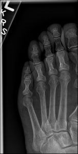
For a fracture, imaging is not just helpful; it is absolutely necessary. For a suspected toe fracture, plain film X-ray is the essential diagnostic tool.
We must obtain high-quality X-rays of the foot and toe. A minimum of two views—like the anteroposterior (AP) and oblique view—is required to visualize the fracture in 3D.
Radiographic features seen in this type of fracture would include:
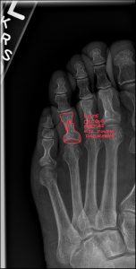
Fracture Line: A visible line of discontinuity in the bone’s structure. For a simple Bedroom Fracture, this might be a fine, hairline crack that is subtle.
Location: The break is clearly seen in the proximal phalanx of the fourth toe.
Fracture Type: The pattern is important—is it transverse, oblique, or perhaps comminuted?
Displacement: Most critically, the image shows whether the bone fragments are in good alignment (non-displaced) or if they have significantly shifted (displaced or angulated). A displaced fracture may require manual reduction.
X-ray: Essential and sufficient. It clearly visualizes the bone break and is the foundation for diagnosis and management.
MRI and CT: Rarely necessary for an isolated, simple phalangeal fracture. They are only considered if there is suspicion of a complex joint injury or a severe, confusing injury pattern.
For a typical Bedroom Fracture, the best practice is to rely on a high-quality X-ray. Advanced imaging is usually reserved for complex scenarios.
MRI (Magnetic Resonance Imaging): Its main use would be in the rare event of a suspected stress fracture not seen on X-ray, or concern for extensive soft tissue damage (like a severe tendon tear) accompanying the bone break.
CT (Computed Tomography): This is reserved for fractures that involve the joint surface (intra-articular fractures). CT provides better 3D visualization of the joint and the amount of “step-off” at the joint, which is information an orthopedic surgeon might need for surgical planning.
Diagnostic Ultrasound: This non-radiation tool is useful for evaluating associated soft tissue injury—like tendon or ligament tears—which can sometimes complicate recovery, even if the bone is healing well.
CBCT (Cone Beam CT): Its role is minimal here, as its best use is for high-detail bone visualization in specific settings, not routine toe fractures.
A Chiropractic radiologist (DACBR) is crucial here, providing Diagnostic Imaging Consultants services. They confirm that the X-ray is sufficient for diagnosis, help the treating chiropractor understand the fracture’s stability, and advise on the rare need for an MRI or CT if the injury proves more complex than a typical chiropractic radiology report might indicate.
The management of an uncomplicated fracture of the proximal phalanx of the fourth toe is primarily conservative.
Referral to Specialists: Referral to a podiatrist or orthopedic surgeon is required if the X-ray, interpreted in the chiropractic radiology report, shows a fracture that is significantly displaced, unstable, or involves the joint surface with a large step-off. These factors increase the risk of a poor outcome and may require manual reduction or surgical fixation.
The Chiropractic Radiologist’s Role: The DACBR ensures the treating chiropractor has the most accurate diagnosis from the imaging. They provide crucial consultation to confirm that the fracture is indeed stable enough for conservative management, thus preventing a malunion that could cause chronic foot problems.
Every day, chiropractors face the same frustration: imaging reports that miss what matters. General radiologists weren’t trained in your world; they don’t understand subluxations, joint dysfunction, or the biomechanical findings that drive your treatment decisions.
The result? Delayed care. Uncertain patients. Cases that stall when they should be progressing.
The Kinetic Radiology Difference: Chiropractors Reading for Chiropractors
Our board-certified DACBRs aren’t just radiologists. We’re chiropractors who chose to specialize in musculoskeletal imaging. We speak your language because we’ve stood where you stand.
Reports You Can Act On Immediately – No vague findings. No irrelevant details. Just the specific insights that guide your next adjustment, your treatment plan, and your patient conversations.
Same-Day Turnaround – Your patients don’t want to wait days wondering what’s wrong. Neither should you. Get clarity fast so care never stalls.
Documentation That Protects Your Practice – Whether it’s insurance requirements, legal protection, or patient records, our reports give you the clinical backing you need.
Confidence That Builds Your Reputation – When patients see you consulting with specialized radiologists, they recognize your commitment to excellence. That trust turns into loyalty, referrals, and five-star reviews.
Think about the last complex case you handled. Did the radiology report actually help you—or did you have to fill in the gaps yourself?
Now imagine having a DACBR partner who catches the subtle findings, flags the red flags, and gives you confidence in every diagnosis.
No commitment. No risk. Just submit your next challenging case and experience what specialized chiropractic radiology can do for your clinical confidence and patient outcomes.
Questions? Call us at 321 325 0096 or email at support@kineticradiology.com
Yes, an X-ray is highly recommended to differentiate a simple contusion or sprain from a true fracture, which requires different management.
While it’s tempting to “tough it out,” symptoms of a simple bruise (contusion) or a ligament sprain can feel very similar to a fracture. However, treating a fracture as a sprain can lead to poor healing, long-term pain, and chronic issues. An X-ray is quick, cheap, and provides the definitive proof needed. The Chiropractic radiologist’s interpretation is crucial here to ensure a small, occult fracture isn’t missed, guiding the chiropractor to apply the appropriate immobilization required for bony healing. The radiation risk from a single toe X-ray is very low compared to the risk of mismanaging a fracture.
The Bedroom Fracture is a simple break of a toe bone (phalanx) from direct impact, while a Jones Fracture is a break of the fifth metatarsal (a long foot bone) caused by twisting, which is notoriously difficult to heal.
Bedroom Fracture is caused by direct trauma, like stubbing a toe, and is generally located in the distal or proximal phalanges. These typically heal well with conservative care. A Jones Fracture occurs further back in the foot, at the base of the fifth metatarsal. This area has a poor blood supply, which makes it prone to non-union (failure to heal) and often requires longer immobilization or even surgery. The Chiropractic radiologist’s job is to carefully review the X-ray to confirm the exact anatomical location and provide this critical differentiation in the chiropractic radiology report.
Yes. If a broken toe heals incorrectly (malunion) or causes prolonged pain, it can permanently alter your walking pattern (gait), leading to secondary problems in the knee, hip, or lower back.
If the pain from the broken toe forces you to limp for weeks, your body compensates. You put more stress on the opposite leg and change the alignment of your pelvis and spine. Even after the toe heals, this altered gait can sometimes become a habit. This is why chiropractic management includes not just fracture stabilization but also gait retraining and adjustments to the spine and pelvis to address these compensatory issues once the bone is stable. Diagnostic Imaging Consultants support the treating chiropractor in ensuring that the original injury heals correctly to prevent these biomechanical issues.
The DACBR (Chiropractic radiologist)’s main role is to provide an expert, formal interpretation of the X-ray.
They generate the detailed chiropractic radiology report, which is essential for the treating chiropractor. This report provides crucial details: the precise location of the fracture, its type (transverse, oblique, etc.), and the degree of displacement or angulation. This information is vital for the treating doctor to decide on the appropriate non-surgical management, or to determine if a prompt referral to a surgeon is necessary for a significantly displaced fracture.
If symptoms strongly suggest a fracture but the initial X-ray is negative, the fracture is considered occult (hidden). The plan usually involves treating the injury as a fracture and getting a follow-up X-ray in 10-14 days.
The second X-ray is often conclusive because the body begins the healing process by reabsorbing bone at the fracture site, making the fracture line clearer. Alternatively, in the case of a suspected stress fracture (a less common Bedroom Fracture cause), an MRI might be ordered to visualize the bone marrow edema that indicates an early break. A Chiropractic radiologist can provide the necessary Diagnostic Imaging Consultants advice on the best course of action.
Partnering with a DACBR teleradiology service provides more than just a second opinion; it offers a significant return on investment:
Speed: Get expert reports in hours, not days.
Expertise: Access board-certified specialists without having to hire them.
Convenience: The entire process is handled online from your office.
Clarity: Receive clear, concise reports that are clinically relevant to chiropractic care, not generic medical reports.
Posted onTrustindex verifies that the original source of the review is Google. Kinetic radiology has been an absolute game changer in speed of reports and detailed reports. Any other doctors I send my reports to are amazed at the detail and the pathology that gets picked up. This is my one and only radiologist group, im thrilled.Posted onTrustindex verifies that the original source of the review is Google. Rishi provides an outstanding service—fast, reliable, and incredibly reassuring. He’s quick to respond, efficient in his work, and always takes the time to address any concerns with clarity and professionalism. I highly recommend his services to anyone looking for a dependable DACBR.Posted onTrustindex verifies that the original source of the review is Google. Prompt efficient service that is thorough and clear. Spinal information is top notch and I've had patients discover kidney stones and possible issues with a hip joint replacement loosening as incidental findings that supported both me and the patient above expectations.Posted onTrustindex verifies that the original source of the review is Google. Quick, accurate, and easy to work with. My new radiology team!Posted onTrustindex verifies that the original source of the review is Google. Excellent, timely reads. Invaluable for CBCTPosted onTrustindex verifies that the original source of the review is Google. Best turnaround time and thorough reports out of any radiologist I’ve seen or worked with!Posted onTrustindex verifies that the original source of the review is Google. Very detailed reports and quick service. Highly recommendedPosted onTrustindex verifies that the original source of the review is Google. Fast turn around time for the radiology reports! Thank you for making this process as seamless as possible!Posted onTrustindex verifies that the original source of the review is Google. I am a NUCCA chiropractor located in Wauankee Wisconsin and I can tell you Dr. Rishi is the only radiologist I’d work with. Sure there are many others in my area but when you want the best you go to the best. He is very easy to work with and always fast to respond and report. 100% recommend.Posted onTrustindex verifies that the original source of the review is Google. Kinetic Radiology is great! They were able to read and get a report written immediately. They are my go to company for any and all images that I need read!Load more
We service all 50 U.S. States, including the following States and Cities listed below.
Copyright 2024 Kinetic Radiology All Rights Reserved
Website Privacy | Terms of UseReceive timely resources to keep you and your practice on the cutting edge of Chiropractic Radiology.
Copyright 2024 Kinetic Radiology
All Rights Reserved
Receive timely resources to keep you and your practice on the cutting edge of Chiropractic Radiology.