A 32-year-old female presents with an insidious onset of left-sided neck, shoulder, and arm pain. She describes the symptoms as a deep ache accompanied by intermittent paresthesia and a feeling of weakness in her hand. Her symptoms are exacerbated by prolonged periods of sitting at her desk for her job as a graphic designer and when performing overhead activities. The condition has been progressively worsening over several months, now interfering with her work and prompting her to seek a definitive diagnosis and care.
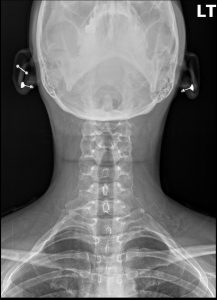
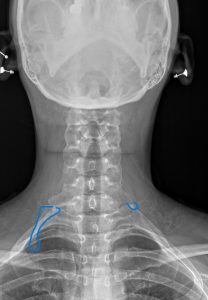
Anteroposterior (AP) and lateral cervical spine radiographs were performed. The AP view clearly demonstrates the presence of bilateral accessory ribs originating from the transverse processes of the C7 vertebra. The left-sided cervical rib is more developed and articulates with the first thoracic rib via a fibrous band, a finding known as a pseudoarthrosis. The remainder of the cervical spine shows preserved vertebral body heights and disc spacing. There is a mild straightening of the expected cervical lordosis, which may be secondary to protective muscle splinting.
Bilateral Cervical Ribs, with anatomical features correlating to clinical symptoms of left-sided neurogenic *Thoracic Outlet Syndrome (TOS)*.
A cervical rib is a congenital anomaly, specifically an accessory rib that arises from the seventh cervical vertebra. The thoracic outlet is the critical anatomical corridor bordered by the anterior scalene muscle, the middle scalene muscle, and the first rib. Through this tight space pass the brachial plexus (the network of nerves supplying the arm) and the subclavian artery and vein. The presence of an anomalous cervical rib can dramatically reduce the dimensions of this outlet, predisposing the neurovascular structures to compression.
Thoracic Outlet Syndrome (TOS) describes the compression of the brachial plexus, subclavian artery, or subclavian vein as they traverse the thoracic outlet. A cervical rib contributes to this compression in two primary ways: first, by directly impinging on the neurovascular bundle, and second, by elevating the floor of the outlet and creating an abnormal fulcrum for the scalene muscles. Chronic compression of the brachial plexus (neurogenic TOS) leads to nerve irritation, resulting in pain, paresthesia, and motor weakness in the upper extremity, matching this patient’s clinical presentation.
While the cervical rib is a congenital condition, it often becomes symptomatic in adulthood due to acquired factors. In this patient, the postural demands of her occupation as a graphic designer—prolonged sitting with forward head posture and elevated arms—likely led to adaptive shortening and hypertrophy of the anterior and middle scalene muscles. This muscular tightening further compromised the already narrowed thoracic outlet, “unmasking” the underlying anatomical variant and initiating the neurovascular compression and subsequent symptoms.
Radiographic features seen in this type of fracture would include:
Identification: An osseous structure arising from the C7 transverse process, seen most clearly on an AP cervical or chest radiograph.
Laterality: May be unilateral or bilateral, but symptoms are often unilateral even when the anomaly is bilateral.
Morphology: Can range from a small, incomplete bony process to a full rib that articulates with the first thoracic rib.
Association: Its presence is a classic and primary cause of true neurogenic TOS.
Plain film radiography is the essential first step for diagnosis. It is a low-cost, low-radiation modality that can definitively confirm or deny the presence of a cervical rib. While X-rays excel at showing bony anatomy, they do not visualize the nerves or vessels directly. In cases where symptoms are severe, progressive, or suggestive of vascular compromise (e.g., arm swelling or discoloration), advanced imaging like MRI or Doppler ultrasound may be warranted to evaluate the brachial plexus and subclavian vessels for direct evidence of compression.
A DACBR provides a crucial service beyond simply spotting the anomaly. The radiologist’s report will characterize the cervical rib’s morphology (e.g., complete vs. incomplete, presence of a pseudoarthrosis), which has clinical implications. By identifying this key anatomical variant, the DACBR provides the treating clinician with a definitive underlying cause for the patient’s complex symptoms. This diagnostic clarity allows the provider to move beyond non-specific neck and arm pain diagnoses and implement a highly targeted treatment plan addressing the specific biomechanics of the thoracic outlet.
Conservative care is the first-line treatment for TOS secondary to a cervical rib and is often very successful. Chiropractic management focuses on relieving compression by addressing the acquired postural and muscular components. This includes manual therapy to release hypertonic scalene and pectoral muscles, specific mobilization of the first rib and cervicothoracic joints, and prescriptive exercises to correct forward head posture and strengthen scapular stabilizers. If a dedicated course of conservative care fails, or if progressive neurologic deficits develop, referral to a vascular or thoracic surgeon for consideration of surgical decompression may be necessary.
Every day, chiropractors face the same frustration: imaging reports that miss what matters. General radiologists weren’t trained in your world; they don’t understand subluxations, joint dysfunction, or the biomechanical findings that drive your treatment decisions.
The result? Delayed care. Uncertain patients. Cases that stall when they should be progressing.
The Kinetic Radiology Difference: Chiropractors Reading for Chiropractors
Our board-certified DACBRs aren’t just radiologists. We’re chiropractors who chose to specialize in musculoskeletal imaging. We speak your language because we’ve stood where you stand.
Reports You Can Act On Immediately – No vague findings. No irrelevant details. Just the specific insights that guide your next adjustment, your treatment plan, and your patient conversations.
Same-Day Turnaround – Your patients don’t want to wait days wondering what’s wrong. Neither should you. Get clarity fast so care never stalls.
Documentation That Protects Your Practice – Whether it’s insurance requirements, legal protection, or patient records, our reports give you the clinical backing you need.
Confidence That Builds Your Reputation – When patients see you consulting with specialized radiologists, they recognize your commitment to excellence. That trust turns into loyalty, referrals, and five-star reviews.
Think about the last complex case you handled. Did the radiology report actually help you—or did you have to fill in the gaps yourself?
Now imagine having a DACBR partner who catches the subtle findings, flags the red flags, and gives you confidence in every diagnosis.
No commitment. No risk. Just submit your next challenging case and experience what specialized chiropractic radiology can do for your clinical confidence and patient outcomes.
Questions? Call us at 321 325 0096 or email at support@kineticradiology.com
Thoracic Outlet Syndrome (TOS) is a condition caused by the compression of nerves, arteries, or veins in the narrow passageway between the collarbone and the first rib, known as the thoracic outlet.
Thoracic Outlet Syndrome (TOS) is a broad term for a group of disorders that occur when the neurovascular bundle—specifically the brachial plexus (nerves), subclavian artery, and subclavian vein—is compressed. This compression happens in a space called the thoracic outlet. This can lead to a range of symptoms in the neck, shoulder, arm, and hand, depending on which structures are being squeezed.
Symptoms typically include pain, numbness, tingling, or weakness in the neck, shoulder, arm, or hand.
The symptoms depend on what is being compressed. The most common form is neurogenic TOS (nerve compression), which causes:
Pain, aching, or throbbing in the neck, shoulder, arm, or hand.
Numbness and tingling (paresthesia), often in the ring and pinky fingers.
Weakened grip strength.
Muscle wasting at the base of the thumb.
If blood vessels are compressed (vascular TOS), symptoms can include a cold, pale hand; arm swelling; or a weak pulse in the arm.
No, most people with cervical ribs have no symptoms at all.
It is estimated that over 90% of individuals with a cervical rib are asymptomatic. The anomaly alone is often not enough to cause a problem. Symptoms typically only develop when another factor—such as a traumatic injury, repetitive overhead motion, or poor posture, it is introduced, leading to compression within the already confined space.
Yes, conservative care like chiropractic and physical therapy is the primary and most effective treatment for the majority of TOS cases.
Absolutely. The goal of this care is to increase the space in the thoracic outlet by addressing the functional components causing compression. A chiropractor or physical therapist will use manual therapy techniques to release tight scalene and pectoral muscles, mobilize the first rib and clavicle, and provide specific exercises to correct posture and improve shoulder mechanics. This approach is often successful in eliminating symptoms without the need for more invasive procedures.
Neurogenic TOS is nerve compression (over 90% of cases) causing pain and tingling, while vascular TOS is artery or vein compression causing swelling, discoloration, or clots.
Neurogenic TOS: The most common type, caused by compression of the brachial plexus. Symptoms are neurological: pain, numbness, tingling, and muscle weakness.
Vascular TOS: A rarer form, subdivided into arterial and venous types. Arterial TOS involves compression of the subclavian artery, causing a cold, pale arm and weak pulse. Venous TOS involves compression of the subclavian vein, leading to arm swelling, blueness (cyanosis), and pain.
Partnering with a DACBR teleradiology service provides more than just a second opinion; it offers a significant return on investment:
Speed: Get expert reports in hours, not days.
Expertise: Access board-certified specialists without having to hire them.
Convenience: The entire process is handled online from your office.
Clarity: Receive clear, concise reports that are clinically relevant to chiropractic care, not generic medical reports.
Posted onTrustindex verifies that the original source of the review is Google. Kinetic radiology has been an absolute game changer in speed of reports and detailed reports. Any other doctors I send my reports to are amazed at the detail and the pathology that gets picked up. This is my one and only radiologist group, im thrilled.Posted onTrustindex verifies that the original source of the review is Google. Rishi provides an outstanding service—fast, reliable, and incredibly reassuring. He’s quick to respond, efficient in his work, and always takes the time to address any concerns with clarity and professionalism. I highly recommend his services to anyone looking for a dependable DACBR.Posted onTrustindex verifies that the original source of the review is Google. Prompt efficient service that is thorough and clear. Spinal information is top notch and I've had patients discover kidney stones and possible issues with a hip joint replacement loosening as incidental findings that supported both me and the patient above expectations.Posted onTrustindex verifies that the original source of the review is Google. Quick, accurate, and easy to work with. My new radiology team!Posted onTrustindex verifies that the original source of the review is Google. Excellent, timely reads. Invaluable for CBCTPosted onTrustindex verifies that the original source of the review is Google. Best turnaround time and thorough reports out of any radiologist I’ve seen or worked with!Posted onTrustindex verifies that the original source of the review is Google. Very detailed reports and quick service. Highly recommendedPosted onTrustindex verifies that the original source of the review is Google. Fast turn around time for the radiology reports! Thank you for making this process as seamless as possible!Posted onTrustindex verifies that the original source of the review is Google. I am a NUCCA chiropractor located in Wauankee Wisconsin and I can tell you Dr. Rishi is the only radiologist I’d work with. Sure there are many others in my area but when you want the best you go to the best. He is very easy to work with and always fast to respond and report. 100% recommend.Posted onTrustindex verifies that the original source of the review is Google. Kinetic Radiology is great! They were able to read and get a report written immediately. They are my go to company for any and all images that I need read!Load more
We service all 50 U.S. States, including the following States and Cities listed below.
Copyright 2024 Kinetic Radiology All Rights Reserved
Website Privacy | Terms of UseReceive timely resources to keep you and your practice on the cutting edge of Chiropractic Radiology.
Copyright 2024 Kinetic Radiology
All Rights Reserved
Receive timely resources to keep you and your practice on the cutting edge of Chiropractic Radiology.