A 34-year-old male presents to the clinic with persistent knee pain that has been progressively worsening over the past three months. The patient describes the pain as deep, aching, and poorly localized, with intensity ranging from moderate to severe. He reports that the discomfort is present at rest but significantly worsens with weight-bearing activities and exercise. The patient has a documented history of sickle cell disease (hemoglobin SS), diagnosed in childhood, with multiple previous pain crises requiring hospitalization. His medical history includes several episodes of acute chest syndrome and chronic anemia managed with hydroxyurea therapy and folic acid supplementation.
On physical examination, the patient demonstrates point tenderness over the distal femoral metaphysis and proximal tibial region. Range of motion testing reveals mild restriction due to pain, though no significant effusion is noted. The patient walks with an antalgic gait favoring the affected limb. Neurovascular examination is intact with normal pulses and sensation. Given his history of sickle cell disease and the clinical presentation, advanced imaging was ordered to evaluate for potential avascular necrosis or bone infarction.
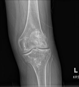
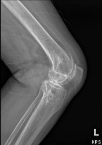
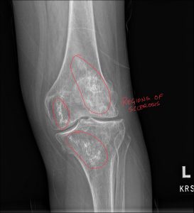
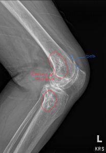
Following comprehensive radiographic and MRI evaluation, the patient was diagnosed with multiple bone infarctions in the distal femur and proximal tibia, consistent with complications of sickle cell disease. The radiology report documented characteristic serpiginous calcifications within the medullary cavity on plain radiographs, along with geographic areas of altered signal intensity on MRI sequences. These findings demonstrated the classic “double line sign” on T2-weighted images, pathognomonic for bone infarction. No evidence of acute osteomyelitis or malignant transformation was identified on imaging studies.
The diagnosis of bone infarction secondary to sickle cell disease was established based on the constellation of clinical history, laboratory values showing chronic hemolytic anemia, and imaging findings characteristic of medullary ischemic necrosis. This case highlights the importance of obtaining detailed radiology reports and, when necessary, seeking a second opinion from specialists with advanced credentials such as DACBR to ensure accurate interpretation of complex osseous pathology.
Understanding bone infarction requires knowledge of osseous vascular supply. Long bones receive blood through several sources:
Vascular Structure | Location | Function | Vulnerability |
Nutrient Artery | Enters through nutrient foramen in diaphysis | Primary blood supply to medullary cavity | Susceptible to thrombosis and embolic events |
Metaphyseal Arteries | Penetrate metaphyseal cortex | Supply metaphyseal region and growth plate | Vulnerable in sickle cell vaso-occlusive crises |
Epiphyseal Arteries | Enter epiphysis | Nourish subchondral bone and articular cartilage | Primary site affected in osteonecrosis |
Periosteal Arteries | Outer cortical surface | Supply outer third of cortex | Generally preserved in bone infarction |
Vascular Occlusion: The initiating event in bone infarction is interruption of medullary blood flow. In sickle cell disease, abnormal hemoglobin polymerization causes red blood cells to assume a rigid, sickle shape. These deformed cells occlude small vessels within the bone marrow, creating areas of ischemia. The relatively slow blood flow and low oxygen tension in the medullary sinusoids make these vessels particularly susceptible to sickling and vascular occlusion.
Ischemic Cascade: Once blood flow is interrupted, bone marrow cells and trabecular osteocytes undergo hypoxic injury and cell death. The ischemic zone develops a central area of complete necrosis surrounded by a border zone of reversibly injured tissue. If blood flow is not restored, the entire region progresses to irreversible necrosis.
Healing Response: Following the acute ischemic event, the body initiates a repair process. Viable tissue at the periphery of the infarct attempts to revascularize the necrotic zone. Dead marrow is gradually replaced by granulation tissue, which subsequently undergoes fibrosis. Calcium salts deposit within the necrotic tissue, creating the characteristic calcifications visible on radiographs. Eventually, the infarcted region may undergo ossification, converting to dense, avascular bone.
Complications: Chronic bone infarctions may weaken bone structure, predisposing to pathologic fractures. Rarely (less than 1% of cases), malignant transformation to sarcoma can occur years or decades after the initial infarction, typically manifesting as osteosarcoma, malignant fibrous histiocytoma, or fibrosarcoma.
Bone infarctions in sickle cell disease typically demonstrate a multifocal distribution, affecting multiple bones simultaneously or sequentially. The distal femur and proximal tibia are most commonly involved, followed by the humerus, fibula, and small bones of the hands and feet. This distribution pattern reflects the anatomic locations with the highest concentration of red marrow and sluggish blood flow.
The clinical presentation of bone infarction varies considerably depending on the acuity, extent, size, and location of the ischemic event.
Presentation | Characteristics | Duration | Associated Findings |
Acute Pain Crisis | Severe, sudden onset pain; poorly localized | Days to weeks | Fever, elevated inflammatory markers, leukocytosis |
Subacute Pain | Moderate, gradually developing discomfort | Weeks to months | Mild functional limitation, antalgic gait |
Chronic Pain | Dull, aching pain; activity-related | Months to years | Progressive functional decline, compensatory patterns |
Asymptomatic | No pain; incidental finding | N/A | Discovered on imaging for other reasons |
Patients with symptomatic bone infarction typically demonstrate:
Local Tenderness: Point tenderness over the affected bone, particularly the metaphyseal regions of long bones. Palpation may reveal warmth and soft tissue swelling in acute cases.
Reduced Range of Motion: If the infarction is located near a joint, patients may exhibit restricted range of motion due to pain or secondary joint stiffness.
Antalgic Gait: Lower extremity involvement often produces a characteristic limping gait pattern where the patient minimizes weight-bearing time on the affected limb.
Muscle Atrophy: Chronic cases may show visible muscle wasting in the affected extremity due to prolonged disuse and pain-related inhibition.
Absence of Inflammatory Signs: Unlike osteomyelitis, bone infarction typically does not produce dramatic erythema, warmth, or fluctuance, though mild soft tissue swelling may be present.
Imaging plays a crucial role in diagnosing bone infarction and differentiating it from other conditions. Working with Diagnostic Imaging Consultants who possess DACBR credentials ensures comprehensive evaluation of subtle findings that may influence diagnosis and management.
Modality | Acute Findings | Chronic Findings | Advantages | Limitations |
Plain Radiography | Often normal initially | Serpiginous calcifications, “smoke-like” densities | Widely available, cost-effective | Low sensitivity in early stages |
MRI | Edema, geographic signal changes, double line sign | Low signal areas with peripheral enhancement | High sensitivity, excellent soft tissue detail | Expensive, time-consuming |
CT | Subtle density changes | Calcifications, ossification, cortical changes | Superior bone detail | Radiation exposure, limited soft tissue contrast |
Bone Scan | Decreased uptake (cold spot) acutely | Variable uptake patterns | High sensitivity for metabolic activity | Poor specificity, radiation exposure |
Radiographic Features: Chronic bone infarctions demonstrate characteristic serpentine or geographic calcifications within the medullary cavity. These calcifications outline the margin between viable and necrotic bone, creating patterns described as “smoke-like,” “lace-like,” or following the contours of the medullary cavity. The cortex typically remains intact unless complications such as pathologic fracture have occurred.
MRI Findings: MRI represents the most sensitive imaging modality for detecting bone infarction. Acute infarctions show diffuse marrow edema pattern on fluid-sensitive sequences. The pathognomonic “double line sign” appears on T2-weighted images, consisting of an inner hypointense line representing sclerotic bone and an outer hyperintense line representing granulation tissue at the interface between viable and necrotic tissue.
A comprehensive radiology report will document the location, extent, age (acute versus chronic), and presence of any complications. This detailed documentation proves essential for treatment planning and longitudinal monitoring.
Accurate diagnosis requires careful consideration of conditions that may mimic bone infarction radiographically or clinically. Additionally, understanding the diverse etiologies of bone infarction is essential for appropriate management and preventing future occurrences.
Etiology | Mechanism | Population Affected | Key Features |
Sickle Cell Disease | RBC sickling causing vascular occlusion | Primarily African descent | Most common cause; multifocal involvement |
Corticosteroid Use | Lipid emboli, marrow fat hypertrophy | Any patient on chronic steroids | Dose and duration dependent |
Alcohol Abuse | Fat emboli, coagulopathy | Chronic heavy drinkers | Often bilateral, symmetric |
Dysbaric Disease (Caisson Disease) | Nitrogen bubble formation | Divers, tunnel workers | Related to rapid decompression |
Gaucher Disease | Marrow infiltration by lipid-laden cells | Ashkenazi Jewish heritage | Erlenmeyer flask deformity |
Pancreatitis | Fat emboli from necrotic pancreas | Acute or chronic pancreatitis patients | Associated with lipase elevation |
Systemic Lupus Erythematosus | Vasculitis, antiphospholipid antibodies | Young to middle-aged women | Part of multisystem disease |
Radiation Therapy | Direct vascular endothelial damage | Cancer patients | Limited to radiation field |
Trauma | Direct vascular injury or fat emboli | Post-fracture patients | Usually adjacent to injury site |
Hyperlipidemia | Fat emboli | Patients with metabolic syndrome | Associated with atherosclerotic disease |
Sickle cell disease represents the most common cause of bone infarction worldwide. The abnormal hemoglobin S polymerizes under hypoxic conditions, causing red blood cells to become rigid and sickle-shaped. These deformed cells occlude small vessels throughout the body, with the bone marrow being particularly vulnerable.
Key Clinical Features:
Other hemoglobinopathies, including thalassemia major and sickle-thalassemia, can also cause bone infarctions, though less frequently than sickle cell disease.
Corticosteroid use represents the second most common cause of bone infarction in developed countries. The risk increases with higher doses and prolonged duration of therapy.
Pathophysiologic Mechanisms:
Risk Factors:
Conditions commonly requiring long-term corticosteroid therapy include systemic lupus erythematosus, rheumatoid arthritis, inflammatory bowel disease, organ transplantation, and asthma.
Chronic alcohol abuse significantly increases the risk of bone infarction through multiple mechanisms including fat emboli, direct toxic effects on bone cells, altered lipid metabolism, coagulopathy, and nutritional deficiencies affecting bone health.
Clinical Patterns:
Decompression sickness affects individuals exposed to rapid decreases in ambient pressure, particularly commercial divers, tunnel workers, and caisson workers. Nitrogen bubbles form in blood and tissues during rapid ascent or decompression, causing vascular occlusion.
Characteristic Features:
This autosomal recessive lysosomal storage disorder results from deficiency of glucocerebrosidase enzyme, leading to accumulation of lipid-laden macrophages (Gaucher cells) in bone marrow and other organs.
Skeletal Manifestations:
Acute pancreatitis can cause bone infarctions through release of fat emboli from necrotic pancreatic tissue and enzymes. This represents a rare but recognized complication.
Associated Features:
Systemic lupus erythematosus (SLE), antiphospholipid syndrome, and other autoimmune conditions increase bone infarction risk through vasculitis, hypercoagulability, antiphospholipid antibodies causing thrombosis, and corticosteroid therapy used for disease management.
Radiation Therapy: Ionizing radiation damages vascular endothelium and bone marrow cells within the treatment field, potentially causing bone infarction months to years later.
Pregnancy: Rare cases of bone infarction during pregnancy or postpartum period, possibly related to hypercoagulable state and hemodynamic changes.
Idiopathic: In approximately 20 to 25% of cases, no clear etiology is identified despite thorough investigation.
Accurate diagnosis requires differentiating bone infarction from other conditions with similar clinical or radiographic presentations.
Condition | Clinical Features | Imaging Characteristics | Age Group | Key Distinguishing Features |
Bone Infarction | Deep bone pain, history of risk factors | Serpiginous medullary calcifications, double line sign | Variable based on etiology | Medullary location, spares subchondral bone |
Osteomyelitis | Fever, warmth, acute pain | Periosteal reaction, cortical destruction, soft tissue changes | Any age | Systemic signs, elevated WBC, positive cultures |
Primary Bone Tumors | Progressive pain, mass, pathologic fracture | Destructive lesion, soft tissue mass, periosteal reaction | Variable by tumor type | Aggressive features, progressive growth |
Bone Metastases | Known primary cancer, multiple lesions | Lytic or blastic lesions, cortical destruction | 50+ typically | History of malignancy, multiple sites |
Enchondroma | Often asymptomatic, incidental finding | Lobulated cartilage matrix, ring and arc calcifications | 20 to 40 | Cartilage matrix, well-defined margins |
Fibrous Dysplasia | Pathologic fracture, deformity, pain | Ground glass matrix, expansile, well-defined | Childhood to young adult | Ground glass appearance, expansile growth |
Paget Disease | Bone pain, deformity, increased hat size | Cortical thickening, trabecular coarsening, mixed lytic/blastic | 55+ | Dramatic bone enlargement, elevated alkaline phosphatase |
Chronic Osteomyelitis | Draining sinus, recurrent infections | Sequestrum, involucrum, sinus tracts | Any age following acute OM | Sequestrum formation, clinical infection |
Osteopetrosis | Pathologic fractures, dense bones | Diffuse increased density, bone-in-bone appearance | Childhood or adult forms | Generalized increased density, multiple bones |
Osteomyelitis represents the most critical differential diagnosis requiring urgent differentiation from bone infarction, particularly in sickle cell patients who have increased susceptibility to Salmonella and Staphylococcus infections.
Distinguishing Features:
Feature | Bone Infarction | Osteomyelitis |
Onset | Gradual or episodic | Acute, rapid progression |
Fever | Absent or low-grade | High fever common |
Laboratory | Normal or mild elevation | Markedly elevated WBC, ESR, CRP |
Blood Cultures | Negative | May be positive |
Imaging Timeline | Changes develop over weeks | Rapid changes over days |
MRI Enhancement | Peripheral rim enhancement | Diffuse marrow and soft tissue enhancement |
Clinical Response | Gradual improvement | Worsens without antibiotics |
In sickle cell patients experiencing bone pain, differentiating between vaso-occlusive crisis with bone infarction versus acute osteomyelitis can be extremely challenging. When uncertainty exists, consulting with Diagnostic Imaging Consultants who hold DACBR credentials provides expert interpretation that may reveal subtle imaging clues favoring one diagnosis over the other.
Several benign and malignant primary bone tumors may produce imaging findings potentially confused with bone infarction.
Enchondroma: This benign cartilage tumor typically occurs in the medullary cavity of short tubular bones (hands and feet) or long bones. Unlike bone infarction, enchondromas demonstrate ring and arc calcifications representing cartilage matrix rather than serpentine calcifications at the infarct margin.
Bone Sarcomas: Primary malignant tumors including osteosarcoma, chondrosarcoma, and Ewing sarcoma can rarely arise within chronic bone infarcts (secondary malignancy) or may be confused with infarction initially. Concerning features include progressive pain, rapid growth, soft tissue mass, aggressive periosteal reaction (Codman triangle, sunburst pattern), and cortical destruction. Any suspicious features warrant prompt biopsy and oncologic evaluation.
Giant Cell Tumor: These benign but locally aggressive tumors occur in the epiphysis extending to the subchondral bone, with a predilection for the distal femur and proximal tibia around the knee. They present as lytic, expansile lesions rather than calcified medullary densities.
Metastatic disease from primary cancers (breast, lung, prostate, kidney, thyroid) commonly affects the skeleton. Multiple lesions, known primary malignancy, age over 50 years, lytic or blastic pattern (rather than serpiginous calcification), and rapid progression help distinguish metastases from bone infarction.
This benign fibro-osseous lesion replaces normal bone with fibrous tissue and woven bone, creating a characteristic “ground glass” matrix on imaging. Unlike bone infarction, fibrous dysplasia typically causes bone expansion, has a predilection for the proximal femur and ribs, and may be associated with McCune-Albright syndrome (polyostotic disease, café-au-lait spots, endocrine abnormalities).
Paget disease involves excessive bone remodeling with accelerated bone turnover. Distinguished from bone infarction by cortical thickening, trabecular coarsening, bone enlargement, involvement of entire bone (rather than focal medullary changes), dramatic elevation of serum alkaline phosphatase, and characteristic imaging patterns (flame-shaped lytic lesions advancing through bone, cotton wool appearance in skull).
This autoinflammatory condition primarily affects children and adolescents, causing recurrent episodes of bone pain and sterile bone lesions. Distinguished by symmetric multifocal involvement, palmoplantar pustulosis or psoriasis, elevated inflammatory markers during flares, absence of organisms on culture, and response to anti-inflammatory therapy.
Given the breadth of potential differential diagnoses and etiologies, comprehensive evaluation by experienced radiologists proves essential. A detailed radiology report should address not only the primary findings consistent with bone infarction but also evaluate for features suggesting alternative diagnoses or complications. When managing complex cases, obtaining a second opinion from specialists in chiropractic radiology or musculoskeletal radiology ensures optimal diagnostic accuracy and appropriate treatment planning.
Intervention | Acute Phase | Subacute/Chronic Phase | Special Considerations |
High-Velocity Manipulation | ❌ Contraindicated | ⚠️ Use extreme caution | Risk of pathologic fracture through weakened bone |
Gentle Mobilization | ⚠️ May be tolerated | ✓ Generally appropriate | Grade I-II techniques, avoid deep pressure |
Active Exercise | 🔶 Modified only | ✓ Recommended | Start with low-impact, progress gradually |
Strengthening | ❌ Avoid initially | ✓ Progressive resistance | Strengthen periarticular muscles for joint protection |
Weight-Bearing | 🔶 Reduce load | ✓ As tolerated | May require assistive devices initially |
Deep Tissue Work | ❌ Contraindicated | ⚠️ Avoid over infarct | Risk of trauma to compromised tissue |
Modalities | ✓ Ice, electrical stim | ✓ Various modalities | For pain management and inflammation |
Approach | Acute Phase | Chronic Phase | Prevention Focus |
Conservative Management | Pain control, rest, activity modification | Gradual return to activity, PT | Address modifiable risk factors |
Medical Management | Analgesics, management of underlying disease | Continue disease-modifying therapy | Optimize treatment of primary condition |
Surgical Intervention | Rarely indicated | Core decompression (controversial), treatment of complications | Prevent pathologic fractures |
Rehabilitation | Gentle ROM, non-weight-bearing exercise | Progressive strengthening, functional training | Maintain bone and joint health |
A DACBR provides a crucial service beyond simply spotting the anomaly. The radiologist’s report will characterize the cervical rib’s morphology (e.g., complete vs. incomplete, presence of a pseudoarthrosis), which has clinical implications. By identifying this key anatomical variant, the DACBR provides the treating clinician with a definitive underlying cause for the patient’s complex symptoms. This diagnostic clarity allows the provider to move beyond non-specific neck and arm pain diagnoses and implement a highly targeted treatment plan addressing the specific biomechanics of the thoracic outlet.
Every day, chiropractors face the same frustration: imaging reports that miss what matters. General radiologists weren’t trained in your world; they don’t understand subluxations, joint dysfunction, or the biomechanical findings that drive your treatment decisions.
The result? Delayed care. Uncertain patients. Cases that stall when they should be progressing.
The Kinetic Radiology Difference: Chiropractors Reading for Chiropractors
Our board-certified DACBRs aren’t just radiologists. We’re chiropractors who chose to specialize in musculoskeletal imaging. We speak your language because we’ve stood where you stand.
Reports You Can Act On Immediately – No vague findings. No irrelevant details. Just the specific insights that guide your next adjustment, your treatment plan, and your patient conversations.
Same-Day Turnaround – Your patients don’t want to wait days wondering what’s wrong. Neither should you. Get clarity fast so care never stalls.
Documentation That Protects Your Practice – Whether it’s insurance requirements, legal protection, or patient records, our reports give you the clinical backing you need.
Confidence That Builds Your Reputation – When patients see you consulting with specialized radiologists, they recognize your commitment to excellence. That trust turns into loyalty, referrals, and five-star reviews.
Think about the last complex case you handled. Did the radiology report actually help you—or did you have to fill in the gaps yourself?
Now imagine having a DACBR partner who catches the subtle findings, flags the red flags, and gives you confidence in every diagnosis.
No commitment. No risk. Just submit your next challenging case and experience what specialized chiropractic radiology can do for your clinical confidence and patient outcomes.
Questions? Call us at 321 325 0096 or email at support@kineticradiology.com
Thoracic Outlet Syndrome (TOS) is a condition caused by the compression of nerves, arteries, or veins in the narrow passageway between the collarbone and the first rib, known as the thoracic outlet.
Thoracic Outlet Syndrome (TOS) is a broad term for a group of disorders that occur when the neurovascular bundle—specifically the brachial plexus (nerves), subclavian artery, and subclavian vein—is compressed. This compression happens in a space called the thoracic outlet. This can lead to a range of symptoms in the neck, shoulder, arm, and hand, depending on which structures are being squeezed.
Symptoms typically include pain, numbness, tingling, or weakness in the neck, shoulder, arm, or hand.
The symptoms depend on what is being compressed. The most common form is neurogenic TOS (nerve compression), which causes:
Pain, aching, or throbbing in the neck, shoulder, arm, or hand.
Numbness and tingling (paresthesia), often in the ring and pinky fingers.
Weakened grip strength.
Muscle wasting at the base of the thumb.
If blood vessels are compressed (vascular TOS), symptoms can include a cold, pale hand; arm swelling; or a weak pulse in the arm.
No, most people with cervical ribs have no symptoms at all.
It is estimated that over 90% of individuals with a cervical rib are asymptomatic. The anomaly alone is often not enough to cause a problem. Symptoms typically only develop when another factor—such as a traumatic injury, repetitive overhead motion, or poor posture, it is introduced, leading to compression within the already confined space.
Yes, conservative care like chiropractic and physical therapy is the primary and most effective treatment for the majority of TOS cases.
Absolutely. The goal of this care is to increase the space in the thoracic outlet by addressing the functional components causing compression. A chiropractor or physical therapist will use manual therapy techniques to release tight scalene and pectoral muscles, mobilize the first rib and clavicle, and provide specific exercises to correct posture and improve shoulder mechanics. This approach is often successful in eliminating symptoms without the need for more invasive procedures.
Neurogenic TOS is nerve compression (over 90% of cases) causing pain and tingling, while vascular TOS is artery or vein compression causing swelling, discoloration, or clots.
Neurogenic TOS: The most common type, caused by compression of the brachial plexus. Symptoms are neurological: pain, numbness, tingling, and muscle weakness.
Vascular TOS: A rarer form, subdivided into arterial and venous types. Arterial TOS involves compression of the subclavian artery, causing a cold, pale arm and weak pulse. Venous TOS involves compression of the subclavian vein, leading to arm swelling, blueness (cyanosis), and pain.
Partnering with a DACBR teleradiology service provides more than just a second opinion; it offers a significant return on investment:
Speed: Get expert reports in hours, not days.
Expertise: Access board-certified specialists without having to hire them.
Convenience: The entire process is handled online from your office.
Clarity: Receive clear, concise reports that are clinically relevant to chiropractic care, not generic medical reports.
Posted onTrustindex verifies that the original source of the review is Google. Kinetic radiology has been an absolute game changer in speed of reports and detailed reports. Any other doctors I send my reports to are amazed at the detail and the pathology that gets picked up. This is my one and only radiologist group, im thrilled.Posted onTrustindex verifies that the original source of the review is Google. Rishi provides an outstanding service—fast, reliable, and incredibly reassuring. He’s quick to respond, efficient in his work, and always takes the time to address any concerns with clarity and professionalism. I highly recommend his services to anyone looking for a dependable DACBR.Posted onTrustindex verifies that the original source of the review is Google. Prompt efficient service that is thorough and clear. Spinal information is top notch and I've had patients discover kidney stones and possible issues with a hip joint replacement loosening as incidental findings that supported both me and the patient above expectations.Posted onTrustindex verifies that the original source of the review is Google. Quick, accurate, and easy to work with. My new radiology team!Posted onTrustindex verifies that the original source of the review is Google. Excellent, timely reads. Invaluable for CBCTPosted onTrustindex verifies that the original source of the review is Google. Best turnaround time and thorough reports out of any radiologist I’ve seen or worked with!Posted onTrustindex verifies that the original source of the review is Google. Very detailed reports and quick service. Highly recommendedPosted onTrustindex verifies that the original source of the review is Google. Fast turn around time for the radiology reports! Thank you for making this process as seamless as possible!Posted onTrustindex verifies that the original source of the review is Google. I am a NUCCA chiropractor located in Wauankee Wisconsin and I can tell you Dr. Rishi is the only radiologist I’d work with. Sure there are many others in my area but when you want the best you go to the best. He is very easy to work with and always fast to respond and report. 100% recommend.Posted onTrustindex verifies that the original source of the review is Google. Kinetic Radiology is great! They were able to read and get a report written immediately. They are my go to company for any and all images that I need read!Load more
We service all 50 U.S. States, including the following States and Cities listed below.
Copyright 2024 Kinetic Radiology All Rights Reserved
Website Privacy | Terms of UseReceive timely resources to keep you and your practice on the cutting edge of Chiropractic Radiology.
Copyright 2024 Kinetic Radiology
All Rights Reserved
Receive timely resources to keep you and your practice on the cutting edge of Chiropractic Radiology.