The ankle joint, known anatomically as the talocrural joint, is a sophisticated synovial hinge joint responsible for the up-and-down motion of the foot (dorsiflexion and plantarflexion). Its remarkable stability comes from a precise bony architecture, often referred to as the ankle “mortise,” which is reinforced by a robust network of ligaments. This mortise is formed by three bones: the tibia, the fibula, and the talus.
Tibia: This is the larger of the two lower leg bones. It forms the roof of the ankle joint (the tibial plafond) and the bony prominence on the inner side of the ankle (the medial malleolus).
Fibula: The thinner, non-weight-bearing bone that runs parallel to the tibia. Its distal end forms the bony prominence on the outer side of the ankle, which is the lateral malleolus.
Talus: This dome-shaped bone sits snugly within the mortise, acting as the crucial connection between the leg and the foot.
The integrity of this bony socket is paramount. It is held together by strong ligaments. On the outer (lateral) side, three main ligaments prevent the ankle from rolling inwards excessively: the anterior talofibular ligament (ATFL), the calcaneofibular ligament (CFL), and the posterior talofibular ligament PTFL). On the inner (medial) side, the powerful deltoid ligament provides stability. Finally, the tibia and fibula are bound together just above the ankle joint by a tough, fibrous structure called the syndesmosis, which is critical for maintaining the width and stability of the mortise.
The pathophysiology of a lateral malleolar fracture is a story of force exceeding the bone’s structural capacity. The most common sequence of events is a supination-inversion injury, where the foot rolls inwards. This places the lateral ligaments under immense tension. The ATFL is usually the first to fail, resulting in a common ankle sprain. If the force continues, it is transmitted directly to the distal fibula. This creates a powerful bending and rotational moment on the bone. The fibula, unable to absorb this acute overload, fractures. The location and pattern of the fracture line are directly related to the position of the foot and the direction of the force at the moment of impact. An unstable fracture occurs when there is associated damage to the medial ligaments (deltoid) or the syndesmosis, causing the talus to shift within the mortise. This loss of congruency is a serious issue that can lead to rapid, debilitating post-traumatic arthritis if not properly addressed.
The mechanism of injury for a lateral malleolar fracture is almost always traumatic and typically involves a forceful twisting or rolling of the ankle. While it can occur from a direct blow to the outer ankle, this is far less common. The vast majority of these injuries result from indirect forces, such as those experienced during sports, falls, or even a simple misstep on uneven ground.
The most classic mechanism is a supination-external rotation (SER) event, which aligns with the Lauge-Hansen classification system. Let’s break down this common sequence:
1. Supination/Inversion: The foot lands and is forced to roll inwards, with the sole of the foot turning towards the midline of the body. This places the lateral ligamentous complex under significant tension. The anterior talofibular ligament (ATFL) is often the first structure to sprain or rupture.
2. External Rotation of the Talus: As the body’s momentum continues forward over the planted foot, the talus rotates externally (outwards) within the ankle mortise.
3. Fracture: This rotational force is transmitted to the fibula, creating an oblique or spiral fracture that typically starts at the level of the joint line and propagates upwards and backwards.
Another common mechanism is a pronation-abduction injury, where the foot is planted and forcibly twisted outwards. This places tension on the medial deltoid ligament first and then creates a compressive force on the lateral side, often resulting in a different fracture pattern, sometimes higher up the fibula.
Understanding the specific mechanism is not just an academic exercise. It helps the clinician predict the likely pattern of injury, anticipate associated ligamentous damage, and determine the overall stability of the ankle. For example, knowing the injury was a high-energy event, like a fall from a height, should raise the index of suspicion for more complex injuries involving the syndesmosis or other bones in the foot and leg. A skilled chiropractor will take a detailed history of the event to piece together these biomechanical clues.
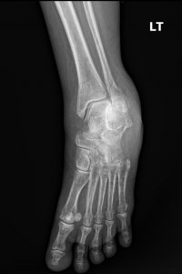
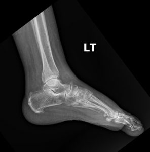
Standard plain film radiography is the cornerstone of diagnosis for a suspected lateral malleolar fracture. A proper ankle series must include at least three views to fully assess the bone and joint integrity:
1. Anteroposterior (AP) View: A straight-on view from the front. It is useful for assessing the general alignment of the tibia and fibula.
2. Lateral View: A side view that is excellent for visualizing the anterior and posterior aspects of the tibia, the talus, and the calcaneus. It is the best view for identifying posterior malleolar fractures and assessing for any anterior or posterior displacement of the talus.
3. Mortise View: This is the most critical view for assessing the stability of the ankle joint. It is taken like an AP view but with the leg internally rotated by 15-20 degrees. This rotation brings the lateral and medial malleoli into the same plane, providing a true, unobstructed view of the entire ankle mortise.
On these images, the radiologist and treating clinician will look for several key features:
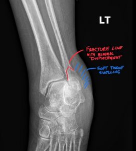
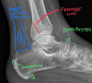
The Fracture Line: Is it visible? Where is it located relative to the joint line (Weber classification A, B, or C)? What is its orientation (transverse, oblique, spiral)? Are the fracture edges sharp (indicating an acute injury) or sclerotic (suggesting an older, non-united fracture)?
Displacement: Are the fracture fragments still aligned, or have they shifted apart? Displacement of more than 2 mm is generally considered significant.
The Medial Clear Space: This is the space between the medial malleolus and the talus, best seen on the mortise view. A space greater than 4-5 mm suggests an injury to the deltoid ligament and indicates ankle instability.
The Tibiofibular Overlap and Clear Space: On the AP and mortise views, there should be a clear overlap between the distal tibia and fibula. A loss of this overlap or a tibiofibular clear space greater than 6 mm suggests a syndesmotic injury (a “high ankle sprain”), which also signifies an unstable ankle.
Talar Tilt or Shift: Is the talus centered within the mortise, or has it shifted sideways or tilted? Any deviation indicates instability.
These meticulous radiographic assessments are what differentiate a simple, stable fracture from a complex, unstable injury that requires a different management approach.
In the modern musculoskeletal healthcare landscape, the role of the board-certified chiropractic radiologist, or DACBR, is indispensable. While many treating chiropractors are proficient at identifying common fractures, the specialized expertise of a DACBR provides a level of diagnostic precision and clinical confidence that is essential for optimal patient care.
When a chiropractor orders an ankle series and sends it to Diagnostic Imaging Consultants for interpretation, they are accessing a specialist who brings a unique focus to the evaluation. The DACBR does more than just spot the fracture line. Their analysis, detailed in the chiropractic radiology reports, focuses on the critical question of joint stability. They will meticulously measure the medial clear space, assess the tibiofibular syndesmosis, and scrutinize the position of the talus within the mortise.
This detailed evaluation is the most important factor in determining the patient’s course of care. A DACBR’s report will explicitly state whether the findings are consistent with a stable or unstable ankle. This provides the treating chiropractor with a definitive answer to the most important clinical question: Can this patient be managed conservatively with immobilization, or do they require an immediate referral to an orthopedic surgeon for surgical fixation?
By providing this clear, evidence-based interpretation, the DACBR empowers the chiropractor to act as a responsible primary spine care provider. It allows them to confidently manage stable fractures within their scope of practice while ensuring that unstable or complex injuries are appropriately triaged to a surgical specialist without delay. This interprofessional collaboration, built on the foundation of expert diagnostic imaging, is the hallmark of modern, patient-centered care.
The treatment plan for a lateral malleolar fracture is dictated entirely by the stability of the ankle.
For Stable, Non-displaced Fractures:
Conservative, non-operative management is the standard of care. The goal is to immobilize the joint to allow the bone to heal in its proper position.
Immobilization: This typically involves placing the patient in a walking boot (cam walker) or a short-leg cast.
Weight-Bearing Status: For the first few weeks, the patient is usually instructed to be non-weight-bearing, using crutches to get around. As healing progresses (confirmed by follow-up X-rays), they are gradually allowed to begin partial and then full weight-bearing in the boot. This entire immobilization phase typically lasts about 6 weeks.
Rehabilitation: This is where a chiropractor or physical therapist plays a leading role. Once immobilization is discontinued, a structured rehabilitation program is crucial. This program focuses on restoring range of motion, rebuilding muscle strength (especially in the peroneal and tibialis muscles), and, most critically, retraining proprioception (the sense of balance and joint position).
For Unstable or Displaced Fractures:
These injuries require an immediate referral to an orthopedic surgeon. Conservative management is not an option, as the ankle will not heal in a stable position, leading to poor function and a high risk of debilitating post-traumatic arthritis.
Surgical Intervention: The standard procedure is an Open Reduction and Internal Fixation (ORIF). The surgeon makes an incision, realigns the bone fragments (Open Reduction), and then fixes them in place with surgical hardware, typically a metal plate and screws (Internal Fixation).
Post-Operative Care: Following surgery, the patient will undergo a similar course of immobilization and progressive rehabilitation as described above, though the timeline may be adjusted by the surgeon. The chiropractor can be an integral part of this post-operative rehab team, collaborating with the surgeon to guide the patient back to full function.
In either scenario, the chiropractor can also provide valuable adjunctive care, addressing secondary issues like gait abnormalities, deconditioning, and compensatory pain in the hip or low back that may arise during the recovery period.
Every day, chiropractors face the same frustration: imaging reports that miss what matters. General radiologists weren’t trained in your world; they don’t understand subluxations, joint dysfunction, or the biomechanical findings that drive your treatment decisions.
The result? Delayed care. Uncertain patients. Cases that stall when they should be progressing.
The Kinetic Radiology Difference: Chiropractors Reading for Chiropractors
Our board-certified DACBRs aren’t just radiologists. We’re chiropractors who chose to specialize in musculoskeletal imaging. We speak your language because we’ve stood where you stand.
Reports You Can Act On Immediately – No vague findings. No irrelevant details. Just the specific insights that guide your next adjustment, your treatment plan, and your patient conversations.
Same-Day Turnaround – Your patients don’t want to wait days wondering what’s wrong. Neither should you. Get clarity fast so care never stalls.
Documentation That Protects Your Practice – Whether it’s insurance requirements, legal protection, or patient records, our reports give you the clinical backing you need.
Confidence That Builds Your Reputation – When patients see you consulting with specialized radiologists, they recognize your commitment to excellence. That trust turns into loyalty, referrals, and five-star reviews.
Think about the last complex case you handled. Did the radiology report actually help you—or did you have to fill in the gaps yourself?
Now imagine having a DACBR partner who catches the subtle findings, flags the red flags, and gives you confidence in every diagnosis.
No commitment. No risk. Just submit your next challenging case and experience what specialized chiropractic radiology can do for your clinical confidence and patient outcomes.
Questions? Call us at 321 325 0096 or email at support@kineticradiology.com
It’s a broken bone on the outside of your ankle.
The lateral malleolus is the end of the fibula bone, which forms the bony bump on the outer side of your ankle. This is one of the most common ankle fractures, usually caused by a twisting or rolling injury.
Only if the fracture is “unstable” or the bone is out of place.
Surgery is reserved for unstable fractures where the ankle joint is no longer aligned. If the fracture is a simple, clean break and the bone fragments haven’t moved (a stable fracture), it can be treated very effectively with a cast or a walking boot to hold it in place while it heals.
The bone takes about 6-8 weeks to heal, but a full recovery and return to sport can take 3-6 months.
While the bone itself mends in about two months, the surrounding muscles, ligaments, and nerves need much more time to recover. A full rehabilitation program is necessary to regain your strength, motion, and balance, which is why the total recovery timeline is significantly longer.
.
It provides the clearest view of the entire ankle joint to check for stability.
A standard X-ray can have bones overlapping, which can hide subtle signs of instability. The mortise view is taken at a special angle that opens up the joint space, allowing the doctor to accurately see if the ankle is properly aligned, which is the most important factor in deciding on treatment.
A fracture is a broken bone, while a sprain is a torn ligament.
A sprain is an injury to the tough, elastic bands (ligaments) that connect bones, whereas a fracture is a break in the bone itself. They can happen at the same time and feel very similar, which is why an X-ray is needed to make a definitive diagnosis.
Partnering with a DACBR teleradiology service provides more than just a second opinion; it offers a significant return on investment:
Speed: Get expert reports in hours, not days.
Expertise: Access board-certified specialists without having to hire them.
Convenience: The entire process is handled online from your office.
Clarity: Receive clear, concise reports that are clinically relevant to chiropractic care, not generic medical reports.
Posted onTrustindex verifies that the original source of the review is Google. Kinetic radiology has been an absolute game changer in speed of reports and detailed reports. Any other doctors I send my reports to are amazed at the detail and the pathology that gets picked up. This is my one and only radiologist group, im thrilled.Posted onTrustindex verifies that the original source of the review is Google. Rishi provides an outstanding service—fast, reliable, and incredibly reassuring. He’s quick to respond, efficient in his work, and always takes the time to address any concerns with clarity and professionalism. I highly recommend his services to anyone looking for a dependable DACBR.Posted onTrustindex verifies that the original source of the review is Google. Prompt efficient service that is thorough and clear. Spinal information is top notch and I've had patients discover kidney stones and possible issues with a hip joint replacement loosening as incidental findings that supported both me and the patient above expectations.Posted onTrustindex verifies that the original source of the review is Google. Quick, accurate, and easy to work with. My new radiology team!Posted onTrustindex verifies that the original source of the review is Google. Excellent, timely reads. Invaluable for CBCTPosted onTrustindex verifies that the original source of the review is Google. Best turnaround time and thorough reports out of any radiologist I’ve seen or worked with!Posted onTrustindex verifies that the original source of the review is Google. Very detailed reports and quick service. Highly recommendedPosted onTrustindex verifies that the original source of the review is Google. Fast turn around time for the radiology reports! Thank you for making this process as seamless as possible!Posted onTrustindex verifies that the original source of the review is Google. I am a NUCCA chiropractor located in Wauankee Wisconsin and I can tell you Dr. Rishi is the only radiologist I’d work with. Sure there are many others in my area but when you want the best you go to the best. He is very easy to work with and always fast to respond and report. 100% recommend.Posted onTrustindex verifies that the original source of the review is Google. Kinetic Radiology is great! They were able to read and get a report written immediately. They are my go to company for any and all images that I need read!Load more
We service all 50 U.S. States, including the following States and Cities listed below.
Copyright 2024 Kinetic Radiology All Rights Reserved
Website Privacy | Terms of UseReceive timely resources to keep you and your practice on the cutting edge of Chiropractic Radiology.
Copyright 2024 Kinetic Radiology
All Rights Reserved
Receive timely resources to keep you and your practice on the cutting edge of Chiropractic Radiology.