A 22-year-old female graduate student with front of the knee pain for the past 5-6 years.
The patient reports first developing knee pain around age 15 while playing competitive volleyball. She was diagnosed with Osgood-Schlatter disease by her pediatrician and told she would “grow out of it.” While the intense pain subsided after she stopped playing volleyball in college, she continues to experience a deep, aching pain directly over the bump on her shin. The pain is exacerbated by activities like kneeling, squatting, or going down stairs. She notes that direct pressure, such as kneeling on the floor to play with her dog, is particularly painful.
Based on the classic clinical presentation and pathognomonic radiographic findings, the diagnosis is Chronic Osgood-Schlatter Disease. For many, it’s a classic “growing pain” of athletic adolescents. But what happens when that pain doesn’t just “go away with age”?
As clinicians, whether you’re a seasoned chiropractor, a physical therapist, or a student just starting your journey, a solid understanding of OSD’s pathophysiology and imaging characteristics is crucial. Accurate diagnosis hinges on great clinical skills and, often, expertly interpreted imaging. That’s where Chiropractic Radiology Experts and board-certified specialists, like a DACBR (Diplomate of the American Chiropractic Board of Radiology), become invaluable partners in patient care. They provide the detailed Radiology Reports that guide our management.
To understand Osgood-Schlatter, we first need to appreciate the elegant, yet vulnerable, anatomy of the adolescent knee. The main players are the quadriceps femoris (the large muscle group on the front of the thigh), the patella (kneecap), the patellar tendon, and the tibial tuberosity.
The quadriceps muscle group is the primary extensor of the knee. It tapers into the quadriceps tendon, which envelops the patella. From the inferior pole of the patella, this structure continues as the patellar tendon (or patellar ligament, depending on your preferred anatomical terminology) and inserts onto a bony prominence on the front of the shin: the tibial tuberosity.
Here’s the critical part for OSD: In a skeletally immature individual, the tibial tuberosity is an apophysis. An apophysis is a secondary ossification center, essentially a piece of growing cartilage that will eventually fuse with the main bone (the tibia). Think of it as a separate piece of bone anchored to the main tibia by a bridge of cartilage (the physis). This apophysis is the direct attachment site for the powerful patellar tendon. This creates a mechanical “tug-of-war” zone, where the immense force of the quadriceps pulls on this relatively weak, cartilaginous anchor.
Osgood-Schlatter disease is not a “disease” in the traditional sense, but rather a traction apophysitis. Let’s break that down:
Traction: A pulling force.
Apophysitis: Inflammation (-itis) of an apophysis.
So, OSD is an overuse injury caused by repetitive strain and micro-avulsions at the insertion of the patellar tendon onto the tibial tuberosity.
This typically occurs during growth spurts in active adolescents, commonly boys aged 10-15 and girls aged 8-13. Activities involving a lot of running, jumping, and sudden changes in direction (think soccer, basketball, gymnastics) place enormous repetitive stress on the quadriceps.
Imagine the patellar tendon as a rope. The quadriceps muscle is a team of strongmen pulling on that rope. The rope is attached to a young, not-yet-solidly-anchored post (the tibial tuberosity apophysis). With every jump and sprint, the strongmen give a mighty heave. Over time, this constant yanking can cause the “post” to become inflamed, irritated, and even sustain tiny fractures where it’s anchored.
The body’s response to this injury is inflammation, which causes the characteristic pain, swelling, and tenderness right over the tibial tuberosity. The body also tries to heal itself by laying down new bone. This healing process, repeated over and over, is what leads to the prominent, bony lump that is the hallmark of OSD.
For about 90% of individuals, the symptoms of OSD resolve once the tibial apophysis fuses with the tibia and skeletal maturity is reached (around age 16-18). However, for a small but significant percentage, the pain persists into adulthood. This brings us to a typical clinical scenario.
Plain film radiography is the first-line imaging modality for suspected Osgood-Schlatter disease. It’s cost-effective, readily available, and excellent for assessing the bony structures. When you send your patient for imaging, the detailed Radiology Reports from Diagnostic Imaging Consultants will describe specific findings that differ between the acute and chronic phases.
In the early stages, radiographs might appear normal. The most common findings, however, are:
Soft Tissue Swelling: Look for swelling anterior to the tibial tuberosity, which may obliterate the sharp definition of the inferior aspect of the patellar tendon.
Thickening of the Patellar Tendon: The distal third of the tendon may appear thickened.
Fragmentation of the Apophysis: The tibial tuberosity apophysis may appear fragmented or irregular. It’s crucial to compare this with the contralateral knee, as ossification can be naturally irregular. However, pronounced fragmentation is a key sign.
Blurring of the Infrapatellar Fat Pad: The normally dark (radiolucent) Hoffa’s fat pad, located just behind the patellar tendon, may show signs of edema, making its lower border appear hazy.
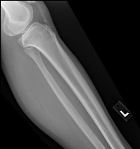
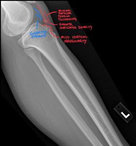
In adults like our 22-year-old patient, the findings reflect the healed, yet altered, state of the anatomy:
Prominent Tibial Tuberosity: The tuberosity itself is often enlarged and knobby due to the heterotopic bone formation during the adolescent healing process.
Intratendinous Ossicle: This is the hallmark of unresolved OSD. It appears as a well-corticated (smooth-bordered) fragment of bone located within the substance of the distal patellar tendon, separate from the tibial tuberosity. This ossicle, sometimes called an “Osgood-Schlatter body,” is essentially a piece of the apophysis that never fused with the main tibia and is now a permanent resident within the tendon. This is the likely pain generator in our case patient, as it can mechanically irritate the tendon and the overlying bursa.
It’s the job of skilled Chiropractic Radiology Experts to differentiate this benign, chronic finding from a more acute avulsion fracture. A DACBR will meticulously analyze the borders of the fragment—smooth and corticated in chronic OSD versus sharp and irregular in an acute fracture.
While X-rays are usually sufficient, sometimes advanced imaging is warranted, especially in chronic or confusing cases.
MRI is the gold standard for evaluating soft tissues and can provide a wealth of information in persistent OSD. An MRI can show:
Patellar Tendinosis: The patellar tendon may appear thickened with abnormal signal intensity, indicating degenerative changes rather than active inflammation.
Bone Marrow Edema: In more active or flared-up chronic cases, there may be bone marrow edema within the tibial tuberosity or the ossicle itself.
Deep and Superficial Infrapatellar Bursitis: The bursae (fluid-filled sacs that reduce friction) around the distal patellar tendon can become inflamed, which is a significant source of pain. MRI is excellent at visualizing this fluid.
Cartilage Integrity: MRI can assess the overlying articular cartilage of the knee joint to rule out other coexisting pathologies like chondromalacia patellae.
Ultrasound is a fantastic, dynamic, and cost-effective tool in the right hands. It provides high-resolution images of the patellar tendon and adjacent structures.
With ultrasound, a clinician can visualize:
Tendon Thickening and Hypoechogenicity: Similar to MRI, this shows signs of tendinosis.
The Ossicle: Ultrasound clearly depicts the bony ossicle and its relationship to the tendon fibers.
Neovascularity: Using color or power Doppler, the sonographer can detect the presence of new blood vessel formation. Neovascularity is often associated with chronic tendon pain and can be a target for certain treatments.
Dynamic Assessment: The clinician can move the patient’s knee during the scan to see if the ossicle is impinging on any surrounding structures. This real-time feedback is a unique advantage of ultrasound.
Both MRI and MSKUS offer a more detailed picture than X-rays, helping to pinpoint the exact pain generator, which is crucial for developing an effective treatment plan.
Treatment strategies for OSD vary significantly between the acute adolescent phase and the chronic adult phase.
The goal here is to reduce pain and inflammation and allow the apophysis to heal without further stress. Management is almost always conservative:
Activity Modification: This is the cornerstone. The athlete must temporarily reduce or cease the offending activities (e.g., jumping, sprinting). Complete rest is rarely needed, but modifying the load is key.
Ice: Applying ice packs for 15-20 minutes after activity can help reduce pain and inflammation.
Stretching and Strengthening: A physical therapy program focusing on stretching the quadriceps and hamstrings, while strengthening the gluteal muscles and core, can help correct biomechanical imbalances that contribute to the strain on the patellar tendon.
Patellar Tendon Straps (Cho-Pat Straps): These straps are worn just below the kneecap. They work by changing the angle of force and distributing the load away from the tibial tuberosity, providing pain relief for many during activity.
For adults like our case patient with a symptomatic ossicle, treatment becomes more nuanced.
Conservative Care First: Many of the same principles apply. A dedicated physical therapy regimen focusing on load management, eccentric strengthening of the quadriceps, and manual therapy can be very effective. Activity modification (e.g., using knee pads for kneeling) is also important.
Injections: For persistent pain associated with inflammation (like bursitis seen on MRI or ultrasound), a corticosteroid injection or a platelet-rich plasma (PRP) injection may be considered to reduce inflammation and promote healing. These should be guided by imaging for accuracy and safety.
Surgical Intervention: In rare, recalcitrant cases where months of dedicated conservative care have failed, surgical excision of the painful ossicle may be an option. The surgery involves removing the free-floating bone fragment from within the tendon. This is typically a last resort but has a high success rate for relieving symptoms in carefully selected patients.
In today’s healthcare landscape, collaboration and specialization are key. By integrating Kinetic Radiology into your practice workflow, you gain a powerful partner that enhances your capabilities and builds your reputation.
Unmatched Expertise: Our reports are generated by board-certified chiropractic radiologists (DACBRs). We are chiropractors who have undergone extensive, multi-year residency training focused solely on musculoskeletal diagnostic imaging. We understand the clinical questions you have because we speak your language.
Speed and Certainty: Don’t wait days for a report. Our streamlined process delivers fast, accurate, and actionable Xray reports, allowing you to make confident clinical decisions in real-time.
A Seamless Extension of Your Practice: Think of us as your in-house radiology department. We provide the expertise you need, when you need it, allowing you to focus on what you do best: treating patients. By ordering an expert report, you demonstrate to your patients that you are committed to the highest standard of care.
For your next complex wrist injury, second opinion, or routine Xray report, choose the experts. Partner with Kinetic Radiology. Contact our team of Diagnostic Imaging Consultants today to see how our DACBR-level insights can benefit your patients and your practice.
Yes, chiropractors effectively manage Osgood-Schlatter by using soft tissue techniques, advising on exercises to correct muscular imbalances, and assessing the entire kinetic chain to reduce stress on the knee.
A chiropractor’s approach to Osgood-Schlatter disease (OSD) is comprehensive and patient-centered. Treatment focuses on alleviating the mechanical stress that causes the condition. This includes performing soft tissue therapies like myofascial release or instrument-assisted techniques on the quadriceps, hamstrings, and hip flexors to reduce tension and improve flexibility. They also provide targeted rehabilitative exercises to strengthen weaker muscle groups, particularly the glutes and core, which helps improve movement patterns and unload the knee joint. Furthermore, chiropractors assess the biomechanics of the entire lower body and spine. By addressing dysfunctions in the feet, hips, or pelvis through adjustments and other modalities,
The most effective conservative care for Osgood-Schlatter is a multi-faceted approach combining activity modification (“relative rest”), icing, a dedicated stretching program for the quadriceps, and a strengthening program for the core and hip muscles.
There is no single magic bullet for OSD; success comes from a consistent, combined strategy. Activity modification is paramount; the patient must reduce or avoid activities that cause pain, like jumping and deep squatting, allowing the inflamed area to heal. This is “relative rest,” not complete immobilization. Icing the tibial tuberosity for 15 minutes after activity is crucial for managing pain and inflammation. A physical therapy or chiropractic rehabilitation program should focus on gently stretching the quadriceps, hamstrings, and hip flexors to decrease the tensile load on the tendon. Critically, this must be paired with strengthening exercises for the gluteal muscles and core. Stronger hips and a stable core improve overall biomechanics, ensuring the quadriceps aren’t overworking and placing excessive strain on the knee during movement. This comprehensive approach addresses both the symptoms and the underlying functional deficits.
A chiropractor orders X-rays to confirm an Osgood-Schlatter diagnosis and, more critically, to rule out other serious pathologies like fractures, bone tumors, or infections that can mimic its symptoms.
While OSD is the most common cause of pain at the tibial tuberosity in active adolescents, it is not the only one. A clinician’s first duty is to ensure an accurate diagnosis and rule out “red flag” conditions. Serious pathologies, although rare, can present with similar localized pain and swelling. These include infections (osteomyelitis), benign tumors (like an osteoid osteoma), and malignant tumors (like osteosarcoma). An X-ray is an essential diagnostic step to visualize the bone and growth plate, confirm the characteristic fragmentation of OSD, and ensure there are no signs of these more aggressive conditions. This imaging provides diagnostic certainty and peace of mind, forming the basis of a safe and appropriate treatment plan. It is a fundamental part of the standard of care.
A Chiropractic Radiologist (DACBR) performs a detailed analysis, looking for soft tissue swelling, thickening of the patellar tendon, and irregular fragmentation of the tibial apophysis in acute cases. In chronic OSD, they identify a persistent, separate ossicle and a prominent tuberosity.
A DACBR provides a specialist’s interpretation that goes beyond simply identifying the main issue. In an acute case of OSD, their report would meticulously detail the degree of anterior soft tissue swelling, irregularity and fragmentation of the secondary ossification center (the apophysis), and any widening of the cartilage space between the apophysis and the tibia. They also assess for subtle signs like haziness in the underlying fat pad (Hoffa’s sign), which indicates inflammation. For a chronic case in an older teen or adult, the DACBR will describe the morphology of the residual ossicle—noting if it’s a single piece or multiple fragments and whether its borders are smooth (indicating chronicity) or sharp (suggesting a recent fracture). This detailed report gives the treating chiropractor a complete picture of the pathology.
Modern teleradiology platforms make getting an expert second opinion simple, fast, and secure.
Find a Chiropractic Teleradiology Service: Choose a reputable service that is staffed by DACBRs. These platforms are designed specifically for chiropractors.
Securely Upload Patient Images: Export the DICOM files from your imaging software and upload them to the service’s secure, HIPAA-compliant online portal. You will also provide a brief clinical history for context.
Receive Your Detailed Report: The DACBR interprets the images and sends a comprehensive, actionable report directly to you, often within 24 hours. This report will clearly state the findings, impressions, and clinical recommendations.
Partnering with a DACBR teleradiology service provides more than just a second opinion; it offers a significant return on investment:
Speed: Get expert reports in hours, not days.
Expertise: Access board-certified specialists without having to hire them.
Convenience: The entire process is handled online from your office.
Clarity: Receive clear, concise reports that are clinically relevant to chiropractic care, not generic medical reports.
Posted onTrustindex verifies that the original source of the review is Google. Kinetic radiology has been an absolute game changer in speed of reports and detailed reports. Any other doctors I send my reports to are amazed at the detail and the pathology that gets picked up. This is my one and only radiologist group, im thrilled.Posted onTrustindex verifies that the original source of the review is Google. Rishi provides an outstanding service—fast, reliable, and incredibly reassuring. He’s quick to respond, efficient in his work, and always takes the time to address any concerns with clarity and professionalism. I highly recommend his services to anyone looking for a dependable DACBR.Posted onTrustindex verifies that the original source of the review is Google. Prompt efficient service that is thorough and clear. Spinal information is top notch and I've had patients discover kidney stones and possible issues with a hip joint replacement loosening as incidental findings that supported both me and the patient above expectations.Posted onTrustindex verifies that the original source of the review is Google. Quick, accurate, and easy to work with. My new radiology team!Posted onTrustindex verifies that the original source of the review is Google. Excellent, timely reads. Invaluable for CBCTPosted onTrustindex verifies that the original source of the review is Google. Best turnaround time and thorough reports out of any radiologist I’ve seen or worked with!Posted onTrustindex verifies that the original source of the review is Google. Very detailed reports and quick service. Highly recommendedPosted onTrustindex verifies that the original source of the review is Google. Fast turn around time for the radiology reports! Thank you for making this process as seamless as possible!Posted onTrustindex verifies that the original source of the review is Google. I am a NUCCA chiropractor located in Wauankee Wisconsin and I can tell you Dr. Rishi is the only radiologist I’d work with. Sure there are many others in my area but when you want the best you go to the best. He is very easy to work with and always fast to respond and report. 100% recommend.Posted onTrustindex verifies that the original source of the review is Google. Kinetic Radiology is great! They were able to read and get a report written immediately. They are my go to company for any and all images that I need read!Load more
We service all 50 U.S. States, including the following States and Cities listed below.
Copyright 2024 Kinetic Radiology All Rights Reserved
Website Privacy | Terms of UseReceive timely resources to keep you and your practice on the cutting edge of Chiropractic Radiology.
Copyright 2024 Kinetic Radiology
All Rights Reserved
Receive timely resources to keep you and your practice on the cutting edge of Chiropractic Radiology.